Review Article
Persistent Napkin Dermatitis: Langerhans Cell Histiocytosis
Murat Durdu1* and Nazım Emrah Koçer2
1Department of Dermatology, Başkent University Faculty of Medicine, Turkey
2Department of Pathology, Başkent University Faculty of Medicine, Turkey
*Corresponding author: Murat Durdu, Department of Dermatology, Başkent University Faculty of Medicine, Adana Hospital, Seyhan, 01250, Adana, Turkey
Published: 08 Jul, 2018
Cite this article as: Durdu M, Koçer NE. Persistent
Napkin Dermatitis: Langerhans Cell
Histiocytosis. Clin Oncol. 2018; 3: 1493.
Keywords
Napkin dermatitis; Langerhans cell histiocytosis; Cytology; Diagnosis
Introduction
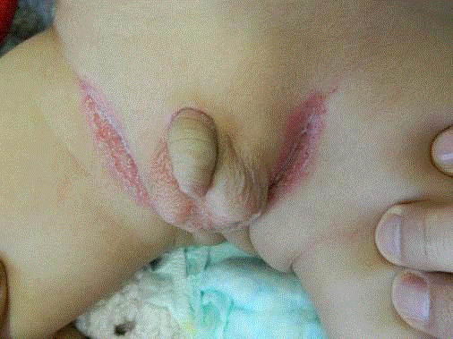
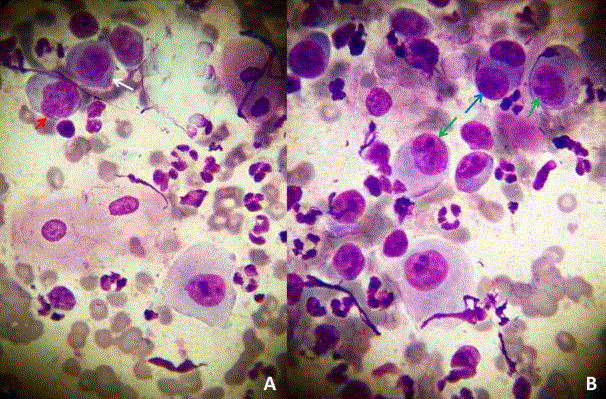
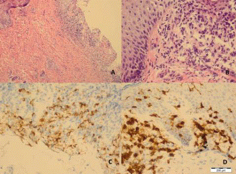
A nine-month-old boy baby was admitted to our dermatology outpatient clinic with persistent napkin dermatitis for three months. Although topical steroid creams were used, these lesions did not improve. On first admission, dermatological examination showed erythematous and erosive inguinal plaques (Figure 1). The other system examination was normal. Potassium Hydroxide (KOH) test was negative. Two separate scraping smears were being taken by scalpel (no: 15). The first sample was stained with May-Grünwald Giemsa stain and the second material was stained with Gram stain. Tzanck smear test showed numerous atypical single or binucleated (blue arrow) histiocytes with eccentric (green arrows), notched (red arrow), vesicular, and grooved nuclei (white arrow) (Figure 2). There was no bacterium in the Gram-stained smear. The histopathology revealed CD1a and Langerin-positive atypical histiocytes compatible with Langerhans Cell Histiocytosis (LCH) (Figure 3). Full blood count, liver function tests, C-reactive protein and bone marrow biopsy were all normal. Chest tomography demonstrated ground-glass opacity. Temporal bone tomography revealed a narrowing of the external auditory canal and lytic lesions of the mastoid bone. Pituitary and brain magnetic resonance imaging, and abdominal ultrasound were normal. These clinical, pathologic and radiographic findings were consistent with Letterer–Siwe disease. Lesions were completely recovered with the combination of vinblastine plus systemic steroid.
Clinical Pearls
Napkin dermatitis is a clinical type of irritant contact dermatitis affects the lower parts of the abdomen, thighs, and the diaper area. Bacteria and/or Candida albicans are also present with these lesions in the chronic stage. The disease should be clinically distinguished from intertrigo, psoriasis, bullous impetigo, acrodermatitis enteropathica, LCH, atopic and seborrheic dermatitis. Napkin dermatitis usually spares the folds, whereas others tend to involve them. The involvement of the other flexural areas should be checked to distinguish napkin dermatitis from seborrheic dermatitis, atopic dermatitis and psoriasis. If the patient has perioral and acral dermatitis, some laboratory tests should be requested in terms of acrodermatitis enteropathica, methylmalonic acidemia. If there is no problem in these laboratory tests, it should be checked for diagnosis of LCH. If hepatosplenomegaly and other systemic symptoms are present, the diagnosis of LCH is not difficult. As in our case, if the initial finding is napkin dermatitis, the diagnosis may be challenging [1]. Clinical expression of LCH ranges from mild, sometimes asymptomatic, single organ involvement to a severe, progressive multisystem disease. LCH represents a disease spectrum with four prominent but overlapping syndromes: Letterer–Siwe disease, Hand– Schüller–Christian disease, eosinophilic granuloma, and congenital self-healing reticulohistiocytosis (Hashimoto-Pritzker disease). Letterer–Siwe disease always develops prior to the age of 2, while Hand–Schüller–Christian disease begins between the ages of 2 and 6 years. Eosinophilic granuloma usually begins between the ages of 7 and 12 years [1].
Figure 1
Figure 2
Figure 2
Tzanck smear test shows numerous atypical single or binucleated
(blue arrow) histiocytes with eccentric (green arrows), notched (red arrow),
vesicular, and grooved nuclei (white arrow) (A and B, May-Grünwald Giemsa
× 1000).
Figure 3
Figure 3
Histopathological examination reveals ulceration of squamous
epithelium due to Langerhans cell infiltration (A). Large magnification
reveals Langerhans cells with large, hyperchormatic, cleaved nuclei (B).
Immunohistochemical examination shows Langerin (C) and CD1a-positive
(D) atypical Langerhans cells in the site of ulceration (A, H&E × 40; B, H&E ×
400; C, Langerin × 200; D, CD1a × 400).
Cytological Pearls
KOH examination should be made to exclude candidiasis in cases of napkin dermatitis. If it is negative, a scraping smear should be taken especially in erosive lesions. Cytological examination does not only show bacteria, but also reveals multinucleated giant cells and acantholytic cells for herpes simplex infections, atypical histiocytes for LCH. If atypical cells are detected in the Tzanck smear examination, histopathological and immunohistochemical studies should be performed [2,3].
Histopathological Pearls
For definitive diagnosis of the LCH, not only histopathological examination but also immunohistochemical examination should be done. Histopathological examination reveals lobule-shaped histiocytes with kidney-like nucleus. The histiocytes have a distinct folded, often kidney-shaped, nucleus. Nucleoli are not prominent, and the slightly eosinophilic cytoplasm is unremarkable. Immunohistochemical examination or electron microscopic examination should be performed for the typing of histiocytosis. In the immunohistochemical examination, Langerhans cells shows positive for S100, CD1a and langerin (CD207). Typical Birbeck granules or Langerhans cell granules can be detected on an electron microscopic examination [4].
References
- Gysel DV. Infections and skin diseases mimicking diaper dermatitis. Int J Dermatol. 2016;55:10-3.
- Ruocco E, Brunetti G, Del Vecchio M, Ruocco V. The practical use of cytology for diagnosis in dermatology. J Eur Acad Dermatol Venereol. 2011;25(2):125-9.
- Ruocco V, Ruocco E. Tzanck smear, an old test for the new millennium: when and how. Int J Dermatol. 1999;38(11):830-4.
- PDQ Pediatric Treatment Editorial Board. Langerhans Cell Histiocytosis Treatment (PDQ®): Health Professional Version. National Cancer Institute (US). 2017.


