Research Article
Investigation of Fiducial Marker Migration during the Treatment of Bladder Cancer with Image-Guided Radiation Therapy
Yulin Song1*, Ming Yan1, Boris Mueller2 and Borys Mychalczak2
1Department of Medical Physics, Memorial Sloan Kettering Cancer Center, USA
2Department of Radiation Oncology, Memorial Sloan Kettering Cancer Center, USA
*Corresponding author: Yulin Song, Department of Medical Physics, Memorial Sloan Kettering Cancer Center Bergen, 225 Summit Ave Montvale, NJ 07645, USA
Published: 10 Jun, 2018
Cite this article as: Song Y, Yan M, Mueller B, Mychalczak
B. Investigation of Fiducial Marker
Migration during the Treatment of
Bladder Cancer with Image-Guided
Radiation Therapy. Clin Oncol. 2018;
3: 1481.
Abstract
Surgically implanted fiducial markers are widely used in the treatment of prostate, lung, and
breast cancers with Image-Guided Radiation Therapy (IGRT). Fiducial markers are typically nonmigrating
gold seeds with various dimensions for different site applications. To prevent markers
from migrating in implanted soft tissue, their surfaces are cross-cut using a special knurling process.
Implanted fiducial markers are felt to constitute a more reliable reference coordinate system than
surrounding bony landmarks for a moving target and are used for point-based image registration.
Studies have shown that fiducial markers are safe, stable, and can provide superior tumor tracking
and targeting capability during both treatment planning and treatment delivery. At our institution,
implanted fiducial markers are now being used for target volume localization in bladder cancer
patients treated with IGRT. In this paper, we present our initial experience with this novel treatment
procedure in terms of fiducial marker migration.
Keywords: Bladder cancer; IGRT; IMRT; OBI; Fiducial marker
Introduction
In 2017, approximately 79,030 Americans were diagnosed with bladder cancer and 16,870 died of this disease [1]. The most common type of bladder cancer is urothelial carcinoma, which accounts for approximately 90% cases in the United States. Based on a 7-year study, the overall 5-year relative survival for all bladder cancer patients was 77.4%. For patients with muscle invasive bladder cancer, excellent local control rates can be achieved with either primary cystectomy or radiation therapy in combination with chemotherapy. However, the majority of the patients with bladder cancer are over 65 years of age, with the median age of 73 at the time of diagnosis [1,2]. For many of these patients, Chemo Radiation Therapy (CRT) is the only safe and curative option. In addition, many younger patients who have had a Complete Response (CR) to induction chemotherapy will prefer bladder preservation therapy. Thus, consolidative CRT is also an attractive treatment option for this group of patients [3,4]. Regardless of the treatment algorithm, delivering high radiation dose safely and accurately to the target in the bladder is a complex process. Due to bladder's anatomical structure and central location in the pelvis, there are unique technical challenges involved in treating bladder cancer using conventional radiation therapy. Firstly, the bladder is a hollow organ with elastic muscular wall. Under normal physiological conditions, its volume undergoes periodic and dramatic variation, from empty to full expansion. The bladder volume change can be as large as 600 ml. This imposes a huge technical challenge on target localization for a safe and accurate radiation delivery. Even though patients are instructed to drink the same amount of water daily prior to each radiation treatment, many have difficulty maintaining a full bladder secondary to irritation by tumor, inflammation related to radiation treatment or due to compression by the immobilization device or mold. Secondly, the position of the bladder relative to the adjacent pelvic bone can be displaced by patient position on the treatment couch. This positioning displacement or organ shift is particularly apparent when the bladder is full. To compensate for these uncertainties in target localization, the most common approach is to expand the Clinical Target Volume (CTV) threedimensionally by a large margin at least equal to the distance change in bladder's radial direction. As a result, the planning target volume (PTV) will include a sizable shell of normal tissue, resulting in unnecessary radiation-induced side effects and damage. Furthermore, the bladder is surrounded by the large and small intestines which are very sensitive to radiation. This is particularly the case for the small intestines, which have a lower dose tolerance. Thus, radiation dose escalation for bladder cancer is challenging without an accurate and reproducible system for target localization in the bladder. The latest generation of medical Linear Accelerators (LINAC) is equipped with an On-Board Imaging System (OBI) in the Kilo-Volts (KV) x-ray range. The modern OBI is capable of producing KV images with a quality close to that of diagnostic x-ray images. However, the low electron density of soft tissue, including bladder tissue, does not exhibit sufficient intrinsic contrast on KV images, which results in poor image contrast. Consequently, target localization based on inherent soft tissue contrast cannot be reliably performed with KV imaging. Although image registration using bony landmarks can be performed using KV imaging, this technique simply cannot track bladder displacement and bladder volume change. An alternative approach to KV imaging would be to use an imaging contrast agent through a catheter during radiation treatment. However, this approach is also impractical because it results in patient discomfort, increases setup time, and poses an infection risk to the patient. In addition, the contrast agent can significantly alter the electron density of the bladder and impact the radiation attenuation coefficient, which can, in turn, affect the delivered radiation dose distribution. Without a reliable image-guidance, radiation therapy in this setting can result in overdosing adjacent critical organs and/or under dosing the target. Over the past few years, surgically implanted fiducial markers have been increasingly used in the treatment of prostate, lung, and breast cancers with Image-Guided Radiation Therapy (IGRT) [5,6]. At our institution, implanted fiducial markers are now being used for target volume localization in bladder cancer patients treated with IGRT. However, unlike other soft tissue sites, such as the prostate and the breast, implanting fiducial markers safely into the thin bladder wall is a more complicated surgical procedure. Our initial assessment of the use of bladder fiducial marker placement is that it significantly improves the accuracy of bladder volume localization and greatly reduces the treatment margin. More importantly, it provides an opportunity for further radiation dose escalation, which may lead to improved tumor control and overall survival. In this paper, we present our initial experience with this novel technique for the treatment of muscle invasive bladder cancer and, in particular, our preliminary assessment of its target localization accuracy and reliability.
Figure 1
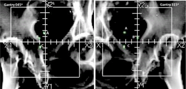
Figure 1
The three implanted fiducial markers are clearly visible on DRRs computed at the gantry angles of 45° (left) and 315° (right). The fiducial markers are
labeled as A, B and C, respectively, and are inside the green contours. One small unit on the digital cross-hair represents 1.0 cm.
Figure 2
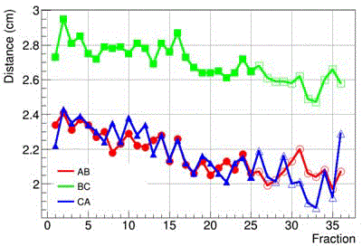
Figure 2
Fiducial marker distances dAB, dBC, and dCA at the gantry angle of
315º as a function of fraction number. Solid markers indicate empty bladder
treatments and hollow markers indicate full bladder treatments.
Figure 3
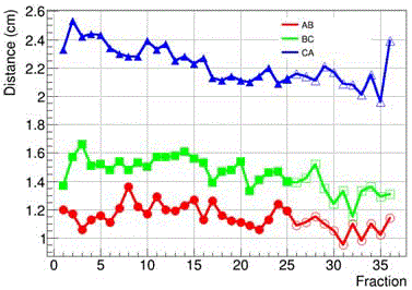
Figure 3
Fiducial marker distances dAB, dBC, and dCA at the gantry angle of
45º as a function of fraction number. Solid markers indicate empty bladder
treatments and hollow markers indicate full bladder treatments.
Materials and Methods
Fiducial Marker Implantation
Our bladder IGRT treatment protocol uses Visicoil gold fiducial
markers (IBA Dosimetry, Bartlett, TN) as the reference points for the
point-based image registration. Visicoil fiducial markers are a helical,
flexible, and thin gold coil. They come with various diameters and
lengths. Usually, smaller diameter markers are optimized for KV and
Cone Beam CT (CBCT) visualization and the larger ones are designed
for Mega-Volts (MV) Electronic Portal Imaging Device (EPID)
visualization. Visicoil fiducial markers are small enough so that they
will not introduce metal streak artifacts on a CT scan and alter the CT
number distribution in the soft tissue near the implantation site [7].
Thus, they are ideal for soft tissue application and IGRT treatment.
Visicoil fiducial markers are pre-loaded on a needle-carrier delivery
device and can be implanted to the patient quickly. For our routine
IGRT treatment, 3 to 4 Visicoil fiducial markers are implanted
into the detrusor muscle wall of the bladder near the site of tumor
resection [8]. Fiducial marker placement is typically performed at
the time of Trans Urethral Resection (TUR), with a needle applicator
passing through a standard adult rigid cystoscope.
IGRT Treatment Planning and Delivery
The patient was simulated and scanned by a Brilliance Big Bore
CT scanner (Philips Medical Systems, Cleveland, OH, USA) in the
supine position one week after the placement of fiducial markers.
The patient was immobilized in a customized thermoplastic mold to
minimize patient movement during the procedure. The patient drank
GoLytely (Braintree Laboratories, Braintree, MA) to empty the bowel
the evening before CT scan. A rectal catheter was placed to localize the
rectum during the scanning procedure. A Foley catheter was not used
for the localization of the urethra. CT images were acquired (3-mm slice
thickness) of the pelvis. An Intensity-Modulated Radiation Therapy
Plan (IMRT) was computed using an in-house treatment planning
system (Top Module). The CTV, urethra, rectum, bowel, and bladder
were delineated on the treatment planning CT scan by a radiation
oncologist. The PTV was created by adding a 1cm margin around the
CTV, except at the interface of the prostate and the rectal wall, where
a 6 mm margin was used. The planner delineated the femoral heads to
include them in the final dose calculation and statistics. In addition,
a series of PTV-concentric tuning structures were also created to
improve the target dose conformity and homogeneity and confine the
dose hot spots to the PTV. Using Boolean operations, the overlapped
structures were optimized independently so that the optimizer could
steer hot spots away from the critical structures. Most treatment plans
consisted of 5 coplanar beams at 225º, 285º, 0º, 75º, and 135º in Varian
IEC scale. Given a set of dose limits and dose–volume constraints,
the plan was optimized by minimizing a quadratic objective function
using an iterative gradient search algorithm. The quadratic objective
function was constructed as the sum of squares of differences between
the desired and actual doses. The algorithm computed the optimal
intensity map for each beam such that the dose distribution resulting
from all beams met the dose constraints specified by the planner. If
the criteria for plan acceptance were not met, a trade-off between
the target dose coverage and the Organ-At-Risk (OAR) constraints
would have to be made. Once optimal intensity maps were obtained,
Multiple-Leaf Collimator (MLC) sequences were subsequently
generated using the Dynamic MLC (DMLC) technique. Based on the
leaf sequences, the final dose distribution was then computed using a
pencil-beam algorithm. During patient treatment, the radiation plan
was delivered on a Varian Trilogy linear accelerator (Varian Medical
Systems, Palo Alto, CA).
Results
In order to investigate the magnitude of migration of the fiducial
markers in the bladder during the course of treatment, we studied
an 84-year old male with bladder cancer recently treated at our
department. This patient was felt to be a good candidate for tracking
the pattern of the fiducial marker migration and assessing the effect
of bladder volume on bladder target localization. The patient had
3 Visicoil fiducial markers implanted in the bladder wall and was
treated with a two-phase IGRT plan: 1) initial phase using 25 fractions
[180 cGy/fx] with an empty bladder, 2) cone down phase using 11
fractions [180 cGy/fx] with a full bladder. The full bladder volume
at the time of phase 2 simulation was 262.6 cc. The Visicoil fiducial
markers implanted into the bladder wall had a diameter 0.35 mm.
The initial distances among those fiducial markers were 3.48, 3.10,
and 2.52 cm, respectively, as measured on the treatment planning CT
scan. Prior to each daily treatment, the patient was setup in reference
to a pair of orthogonal Digitally Reconstructed Radiographs (DRR) at
the gantry angles of 45º and 315º, respectively (Figure 1). The KV OBI
was set at 50 cm away from the MV radiation isocenter. The accuracy
of the patient setup was evaluated by registering the 3 fiducial markers
on the orthogonal pair of radiographic images acquired by the KV
OBI with those on the DRRs. The image registration was performed
using a rigid body model, which yielded three translational shifts
and one rotational shift (yaw). The patient shifts were corrected by
automatically moving the treatment couch to the target position.
Following the shift correction, a new pair of KV images was then
acquired to confirm the patient setup to be within our institutional
tolerance (≤1.0 mm). After a satisfactory patient setup had been
achieved, the radiation treatment was delivered. Post-treatment,
the daily KV images were analyzed off-line using the Varian image
registration software, Offline Review. To facilitate our analysis, we
defined the three fiducial markers as A, B, and C, respectively, and
measured the baseline distance between each pair of the fiducial
markers on the DRRs obtained on the simulation date. The baseline
distances were dAB=2.34cm, dBC=2.73cm, and dCA=2.22cm at the
gantry angle of 315° and dAB=1.2cm, dBC=1.37cm, and dCA=2.33cm at
the gantry angle of 45°. These were the distances projected onto the
isocenter plane as viewed by the MV radiation source and, therefore,
were gantry angle-dependent. To assess the fiducial marker migration
as a function of time, we also measured the distance between each
pair of the fiducial markers on the daily KV images for each treatment
fraction. (Figure 2) shows the distances as measured on daily KV
mages at the gantry angle of 315º as a function of treatment fraction
number. (Figure 3) shows the distances as a function of treatment
fraction number at the gantry angle of 45o. Solid markers on the plots
indicate treatment fractions with empty bladder (phase 1) and hollow
makers indicate those treated with full bladder (phase 2). As seen in
the plots, despite the significant daily bladder volume changes, the
distances between fiducial markers were not drastically changed.
When the plots were fitted to a linear trend line, we found that the
distances decreased at a very small rate of approximately 0.010cm/
fx. In specific, the respective rates were rAB= 0.008 ± 0.001cm/fx,
rBC=-0.011 ± 0.002cm/fx, and rCA=-0.009 ± 0.001cm/fx at the gantry
angle of 315º. The corresponding values at the gantry angle of 45º
were rAB= 0.004 ± 0.001cm/fx, rBC=-0.008 ± 0.001cm/fx, and rCA=-
0.010 ± 0.001cm/fx. When examining the full and empty bladder data
together, we found that the mean distances were dAB=2.16 ± 0.12cm,
dBC=2.70 ± 0.11cm, and dCA=2.17 ± 0.25cm at the gantry angle of 315º
and dAB=1.17 ± 0.09cm, dBC=1.49 ± 0.11cm, and dCA=2.27 ± 0.13cm
at the gantry angle of 45º. They did not drift greatly from the baseline
values. These findings were consistent with the results reported
previously for prostate patients.
Compared to the magnitude of the fiducial marker migration, the
daily couch shifts necessary to correct the random patient setup errors
and the bladder displacement were much larger. For this patient, the
mean daily couch shifts in the Vertical (VRT), Longitudinal (LNG),
and Lateral (LAT) directions were CVRT=0.07 ± 0.35cm/fx, CLNG=0.56
± 0.30cm/fx, and CLAT=-0.39 ± 0.36cm/fx, respectively, for the empty
bladder treatments and CVRT=-0.27 ± 0.28cm/fx, CLNG=1.21 ± 0.41cm/
fx, and CLAT=-0.20 ± 0.43cm/fx for the full bladder treatments (Figure
4). These findings demonstrate that bladder target localization using
implanted fiducial markers is reliable because the coordinate system
established by the fiducial markers remains relatively fixed in space
as compared to the magnitude of the random patient setup errors
and the bladder displacement in the pelvis. We also performed
statistical analysis of the fiducial marker migration. Figure 5 shows
the histogram of the fiducial marker migration dCA as an example.
It followed a normal distribution with a mean of 2.20 cm and the
standard deviation of 0.14 cm. The mean was effectively equal to the
dCA value (2.22 cm) measured at the date of patient simulation. For
about 95% of the treatment fractions, dCA fell between 2.1 and 2.4 cm.
Figure 4
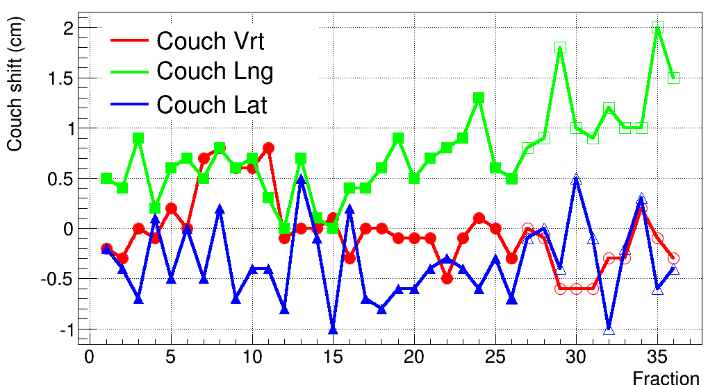
Figure 4
Couch shifts as a function of treatment fraction number. Solid
markers indicate empty bladder treatments and hollow markers indicate full
bladder treatments.
Figure 5
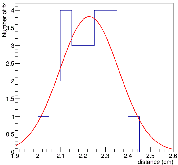
Figure 5
Distribution of fiducial marker migration dCA.
Conclusions
Visicoil fiducial markers have been found to be stable in prostate cases. Several studies have shown that the typical marker migration over an entire treatment course is about 1.0 mm. Similar investigations have been performed in lung, breast, and GI cases. However, bladder Visicoil fiducial marker implantation is a relatively new clinical application and the fiducial marker migration in the bladder has not been fully investigated. Our analysis shows that the distances between two neighboring fiducial markers implanted in the bladder shrank at a mean rate of -0.043±0.082mm per fraction and were not correlated with the bladder volume change. The typical numerical order of fiducial marker migration for the entire treatment course was about 1.0 mm, which was much smaller than the daily couch shifts. This provides a concrete evidence for validating this procedure as a viable IGRT technique in bladder cancer treatment. The use of Visicoil as fiducial markers in bladder cancer patients for IGRT treatment is still a new technique. Implantation of Visicoil fiducial markers into a thin and dynamic bladder wall requires significant clinical experience and extensive training. Currently, the number of bladder cancer patients implanted with Visicoil fiducial markers for IGRT treatment is very limited. More patient data using the bladder Visicoil fiducial markers need to be analyzed to ensure a better understanding of the causes and patterns of fiducial marker migration.
Acknowledgements
This research was funded in part by the NIH/NCI Cancer Center Support Grant P30 CA008748.
References
- National Cancer Institute (NCI), Surveillance, Epidemiology, and End Results Program Cancer Statistics 2017, SEER Stat Fact Sheets.
- Plataniotis G, Dale R. "Radio-chemotherapy for bladder cancer: Contribution of chemotherapy on local control”. World J Radiol. 2013;5(8):267-74.
- Shipley W, Winter K, Kaufman D, Lee W, Heney N, Tester W, et al. Phase III trial of neoadjuvant chemotherapy in patients with invasive bladder cancer treated with selective bladder preservation by combined radiation therapy and chemotherapy: initial results of Radiation Therapy Oncology Group 89-03. J Clin Oncol. 1998;16(11):3576-83.
- Shipley W, Kaufman D, Zehr E, Heney N, Lane SC, Thakral HK, et al. Selective bladder preservation by combined modality protocol treatment: long-term outcomes of 190 patients with invasive bladder cancer. Urology. 2002;60(1):62-7.
- Gates LL, Gladstone DJ, Kasibatla MS, Marshall JF, Seigne JD, Hug E, et al. Prostate localization using serrated gold coil markers. International Journal of Radiation Oncology. 2007;69(3):S382.
- Kupelian PA, Forbes A, Willoughby TR, Wallace K, Mañon RR, Meeks SL, et al. Implantation and stability of metallic fiducials within pulmonary lesions. Int J Radiat Oncol Biol Phys. 2007;69(3):777-85.
- Brook O, Gourtsoyianni S, Brook A, Mahadevan A, Wilcox C, Raptopoulos V. Spectral CT with metal artifacts reduction software for improvement of tumor visibility in the vicinity of gold fiducial markers. Radiology. 2012;263(3):696-705.
- Kollmeier M, Bochner B. Intravesical fiducial marker placement to facilitate image-guided radiation therapy for patients with muscle-invasive bladder cancer. UroToday International Journal. 2011;4(1).