Mini Review
An Interesting Pulmonary Contusion Case
Nese Dursunoglu* and Mukaddes Kılınç
Sleep Laboratory, Pamukkale University Medical Faculty, Denizli, Turkey
*Corresponding author: Nese Dursunoglu, Sleep Laboratory, Pamukkale University Medical Faculty, Denizli, Turkey
Published: 11 May, 2018
Cite this article as: Dursunoglu N, Kılınç M. An Interesting
Pulmonary Contusion Case. Clin Oncol.
2018; 3: 1468.
Introduction
“Pulmonercontusion” is a term that indicates the destruction in lungs after a traumatic accident
without any laceration. It is seen in all age groups; especially children have contusions limited to
lungs because of high mobility of mediastinal tissue and flexibility of thoracic ribs without any
fractures [1].
In this case we present a patient who came with thoracic pain. We realized the pulmonary
contusion after taking a detailed history of patient.
Case
A 65 year old male, obese farmer with hypertension and obstructive sleep apnea syndrome and
taking Angiotensin Converting Enzyme inhibitor (ACE), Diuretics and CPAP (Continuous airway
pressure) at nights. No smoking history.
He came into clinic as an outpatient with his postero-anteriorgraphy (PA-Graphy). He had a
density area which was remaining constant although he had been taking many antibiotics due to
pneumonia diagnosis at left-inferior zone on PA-Graphy. He had no respiratory complaints. There
was not any pathological sign at physical examination (Figure 1).
The vital signs were normal. Oxygen saturation was 99%. Laboratory was also normal.
On thorax Computed Tomography (CT) scan, “Traction bronchiectasis, the fissural contracts
on left inferior lob and central nodules, ground glass sign areas on left lateral basal segment” were
seen (Figure 2 and 3). Despite of broad spectrum anti biotherapy for 2 weeks there had been no
changes on PA-Graphy.
After we questioned the patient history deeply, the patient
remembered that he had been kicked by two cows in his barn. After
that he had a chest pain but it was dissolved in a few days. A week after
the event the pain started again and he applied to clinics and forgot to
mention trauma. He was followed –up without treatment.
Figure 1
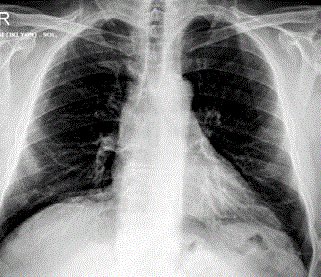
Figure 1
PA- Graphy one year old.
Figure 2
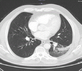
Figure 2
Figure 3
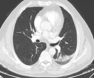
Figure 3
Conclusion
Many of radiologic signs can be noticeable at pulmonary
contusions. For example hemorrhage, consolidations and oedema
Intra alveolar hemorrhages and interstitial oedema are seen especially
in small traumas. Mucosal secretions gather, bronchial airways fillup
with fluid and blood in adjacent lung paranchyma. Atelectasis
and consolidations are building after surfactant production and
compliance decrease, capillary permeability increases [2].
Pulmonary contusions are sometimes misdiagnosed as
pneumonia or malignancy when anamnesis does not take thoroughly.
It may be time consuming and expensive. Therefore we emphasize the
importance of an amnesis to reach the correct diagnosis.