Research Article
Magnetic Resonance Lymphangiography: Clinical and Radiological Correlation
Savaş Tepe*, Ali Ertan Çapar and Ali Rıza Ercocen
Bayindir Hospital, İstanbul, İçerenköy, Turkey
*Corresponding author: Savaş Tepe, Bayindir Hospital, Department of Radiology Bayindir Hospital, Bayindir Hastanesi Ali Nihat Tarlan Cd. Ertas Sk. No:17 Icerenköy, Atasehir, Istanbul, Turkey
Published: 31 Oct, 2017
Cite this article as: Tepe S, Çapar AE, Ercocen
AR. Magnetic Resonance
Lymphangiography: Clinical and
Radiological Correlation. Clin Oncol.
2017; 2: 1362.
Abstract
Aim of this study is to search the correlation between the clinical and pre and post operative findings and three dimensional Magnetic Resonance Lymphangiography (MRL) images of the lower extremities. Total of 10 patients with primary and secondary lymphedema of the lower legs (2 males, 8 females, range 15-80, mean age of 36) were retrospectively evaluated, challenges and technique of MRL were reviewed in patients with lower extremity lymphedema. Demonstration of lymphatics, venules, lymph nodes and surgical lymphaticovenular anastomosis is a lately utilized radiological method which diagnoses presence, extent, intensity of lymphedema; maps and identifies lymphatic vessels; and guides for surgical planning. MRL maintains volumetric datasets that are high in resolution to determine the existence and intensity of lymphedema; illustrates superficial lymphatic vessels; provides anatomic and morphologic information. All our patients were referred by the Aesthetic & Reconstructive Surgery Department. In close relationship we have discussed the results of pre and post operative versions of MRL with them and finally prepared an MRL report that would fulfil their needs. Value of the radiology report for the surgeon in an MRL examination is also emphasized.
Introduction
Purposes of this study are demonstration of lymphatics using Magnetic Resonance Imaging
(MRI) as a guide, seeing the correlation of clinical findings and helping surgeon select appropriate
microsurgical techniques and treatment for lymphedema.
Stages of lymphedema are latent (subclinical), mild, moderate and severe. Latent and mild stages
could respond to conservative approach such as limb elevation and gradient compression garments.
In our study MRL was performed for the patients with severe stages of lymphedema.
Identification or visualization of the lymphatics has a long and remarkable historical challenge.
Lymphedema is a debilitating disease caused by abnormal lymphatic flow and generally associated
with malignancy and also its treatment. Lymphedema is basically described with the following;
inflammation, abnormally gathered protein rich fluid, interstitial space fibrosis, and hypertrophy
of the adipose tissue [1]. Lymphedema is categorized as primary (congenital) or secondary.
Congenital lymphedema is less common compared to secondary which might arise from blockage of
lymph vessels due to operation, trauma, infection or radiation. Breast cancer and gynecological
malignancies and related surgeries are the most frequent malignancies causing secondary
lymphedema [2,3].
Patients with lymphedema secondary to connective tissue diseases, infection and recurrent
cellulitis are not included in this study.
Newer imaging technique with high resolution dynamic three dimensional (3D) MRL,
demonstration of lymphatics, venules, lymph nodes and surgical lymphaticovenular anastomosis is a
lately utilized radiological method which diagnoses presence, extent, intensity of lymphedema, maps
and identifies lymphatic vessels, and guides for surgical planning. MRL maintains volumetric datasets
that are high in resolution to determine the existence and intensity of lymphedema; illustrates
superficial lymphatic vessels; provides anatomic and morphologic information which may differ
chronic phase of the disease.
Materials and Methods
From January 2013 to August 2017 ten patients were evaluated with MRL. Informed consent
was obtained from all individual participants included in the study. The research was performed
according to the Declaration of Helsinki principles. MRL was
applied on a 1.5 Tesla MR imaging magnet (Siemens, Magnetom
Avanto, Erlangen, Germany) employing phased array surface coils.
Imaging protocol used for the MRL, image post processing and
interpretation of the images in our institution are as revealed. Upon
clinical request MRI of the lower extremities was applied bilaterally.
In the MRI magnet patients are laid feet first and lying on their backs,
having the face upward. Surface coils are placed from mid foot to hip.
Head coils are positioned to ankle region. First, high T2 weighted 3D
sequence was used for defining the intensity and scope of lymphedema.
Then intracutaneous contrast medium injected through both feet
interphalangeal area 4 sites each to detect lymph vessels using 3D
gradient echo (GRE) sequence images.
Imaging parameters are as follows;
Pre contrast T2W Fat suppressed coronal plan, repetition time
(TR) 2000 msn, echo time (TE) 693 msn, Flip Angle (FA) 150, field of
view (FOV) 450 mm, section thickness 1 mm, matrix 259 x 320, scan
time 6.5 minutes.
Post contrast T1 spoiled gradient echo (SPGR) fat saturated
coronal plan, TR 4.66, TE 2.38, FA 25, FOV 450, section thickness 1.5
mm, matrix 448 x 448, scan time 2.4 minutes.
First EMLA (Astrazeneca, France) anesthetic cream is put on the
foot distal intermetatarseal- interphalengeal area prior to injection.
The contrast material is prepared while pre contrast scan acquisition
is taken. During intracutaneous contrast administration, patients felt
mild pain. There were no other discomfort or complications related to
study. If there is a case of patient expressing pain and distention, we
recommend dealing with discontent similar to contrast extravasation
which may be seen after intravenous contrast injections (get vitals,
evaluate for compartment syndrome tissue necrosis, raise extremity,
make cold compress, monitor, advise the patient with instructions to
follow additional medical care if symptoms worsen, consult surgery,
report to the patient's physician, note down in the medical record).
Combination of the subcutaneous injection of 2% citanest
(Zenica Medical, Paris, France) 5 cc, 0.1 mmol/kg body weight
gadolinium (multihance, gadobenate dimeglubine, Guerbet, France)
injected to the each interdigital web space between the metatarsals
approximately 2 cc each interdigital web space with a 24 G needle.
The injected sites were massaged for a minute. Acquisition was
done at 5, 15, 30, 45, 60 minutes, 2 hours, 4 hours, 8 and 24 hours.
Following contrast administration venous enhancement always
occurs. Lymphatics enhancement usually augments and slowly
advances with time, whereas enhancement of the veins lessens with
time, therefore kinetic of the enhancement of lymphatics versus veins
are helpful to differentiate.
Enhanced lymphatic channels may not be detected in
standard extremities with MRL which is assumed to be related with
quicker lymphatic transport in a normal extremity.
Table

Table
Table of the patients with lower extremity lymphedema.
Figure 1
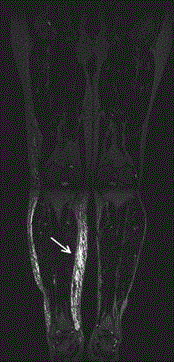
Figure 1
Pre contrast coronal T2W MR image characteristically demonstrates
muscle sparing epifascial distribution of lymphedema (arrow).
Results and Discussion
Patients
Table: Table of the patients with lower extremity lymphedema.
Before administration of the contrast agent, first pre contrast
T2W magnetic resonance images are evaluated. This will provide the
knowledge of distribution of lymphedema (Figure 1). Following this
sequence mapping of lymphatics are in order. Starting at 5 minutes,
followed by 15, 30, 45, 60 minutes, 2 hours, 4 hours, 8 and 24 hours
post contrast magnetic resonance lymphangiography sequences are
performed (Figure 2 and 3).
Dermal back flow is an area of progressive interstitial dispersion
of the contrast medium in soft tissue due to proximal obstruction of
lymph drainage or in another term poor lymphatic drainage, reflects
proximal lymphatic obstruction (Figure 4).
Lymphatico venular anastomosis is performed when finding a
vein in suprafascial area without venous insufficiency, neighboring
lymphatics near and well mapped (Figure 5).
Lymphaticovenous anastomosis redirects a lymphatic obstruction
by conducting distal lymph flow into neighboring veins therefore is
generally performed to treat lymphedema. For the regimen and to
appropriately treat lymphedema, visualization of the lymphatic
channels prior to surgery is important. New dedicated MR imaging
sequences are able to demonstrate lymphatic channels with MRL
and thus help the surgeon plan adequate microsurgery, currently
lymphaticovenular anastomosis and lymph node relocation to
nurture lymphangiogenesis, improve lymphatic drainage, reduce
limb diameter, and avoid dermal sclerosis (Figure 6).
It is also possible to compare this method with other visualization
techniques and methods. Available alternative imaging techniques for
evaluation of lymphedema are bioelectric impedance spectroscopy,
nuclear medicine lymphoscintigraphy and indocyanine green
lymphography. Bioelectric impedance spectroscopy applies
electrical impedance to weight the magnitude of extracellular water
in an extremity. Nuclear Medicine Lymphoscintigraphy (NML)
is frequently used method providing affirmation of unorthodox
lymphatic flow to analyze lymphedema. Lymphatic dysfunction is
identified as slowed asymmetric or non visualization of regional lymph
nodes, unsymmetrical lymphatic channels or dermal back flow. NML
may not portray individual lymphatic channels since it suffers from
poor spatial and temporal resolution [4]. Fluorescent indocyanine
green is infused intracutaneously into the extremities for indocyanine
green lymphography (ICGL) technique, and a photoelectric apparatus
is utilized to detect the indocyanine green fluorescence within the
superficial lymphatic routes and at places of dermal back flow. ICGL
has a limited penetration depth of approximately 2 cm which is short
in depth. The patient is not regarded suitable for reconstruction
with LVA and becomes a possible nominee for excisional surgery or
cosmetic reduction if non functioning superficial lymphatic channels
are detected with MRL.
Figure 2
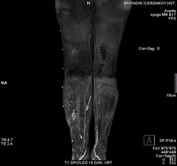
Figure 2
Early visualization of lymphatics at 15 minutes showing lymphatic
networks, discontinuous, tortuous, beaded lymphatic vessels, whereas
veins are smooth and uniform in caliber, linear and continuous (short arrow
lymphatic vessels, long arrow veins).
Figure 3
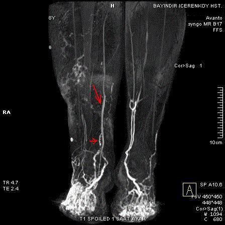
Figure 3
Same patient at 60 minutes (short arrow lymphatic vessels, long
arrow veins).
Figure 4
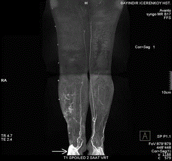
Figure 4
MRL at 2 hours, showing sites of dermal back flow (an area of
progressive interstitial dispersion of the contrast medium in soft tissue due
to proximal obstruction of lymph drainage or in another term poor lymphatic
drainage, reflects proximal lymphatic obstruction ), patchy, non regular high
signal intensities on contrast enhanced image (arrow).
Figure 5
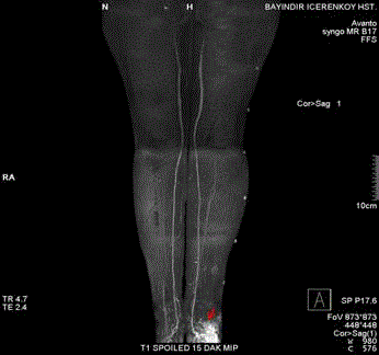
Figure 5
A region of blush (arrow) represents a site of LVA.
Figure 6
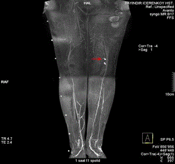
Figure 6
MRL image at 1 hour showing transferred lymph node. (arrow).
Conclusion
In our MRL method we had acquisitions performed at 5, 15, 30,
45, 60 minutes, 2 hours, 4 hours, 8 and 24 hours, in order to evaluate
if there are improvements or benefits examining the patients at
those late intervals. However, this method is demanding and time
consuming. Our findings reveal no great benefit performing the
scanning after 4 hours. Even it is possible to get the most of necessary
information in the first two hours scanning of MRL therefore it will
not add too much extra information in the extended hours despite the
efforts and time spent. If time limitation is an issue, MRL examination
even only the calf lymphatic vessels may be informative. Hence Lu [3]
and colleagues mention that there was insignificant variance when
normal and affected thighs are compared but there was a meaningful
variance in transverse width and numbers of lymphatic channels
between healthy and abnormal calf. They also imply that lymphatic
neoperfusion or neovascularization appears more often in the
abnormal calf than the abnormal thigh. This observation interestingly
is about secondary LEL. On the other hand people with congenital or
praecox types of primary lymphedema types are described as having
hypoplastic lymph routes in the thigh and calf.
We performed MRI sequences in coronal plan for both legs at
the same time. Some centers perform scan orientation in sagittal plan
then transform the images at workstations to Maximum intensity
projections to coronal plan. This may provide better visualization,
however it is not possible to perform MRL for both legs at the same
time and MRL should be performed separately unilaterally for each
leg if MRL would be performed bilaterally in sagittal plan. The patient
should be centered to the magnet, and the legs should be placed
as nearly as possible to the scanner isocenter. This helps promote
shimming and homogeneity of fatty tissue elimination.
Some limitations of the MRL must be mentioned and these are
long duration of the MR examination, and infrequent difficulty in
characterizing the affected lymphatic vessels when an underlying
venous contamination is present. Pelvic and above knee region were
insufficient to image for lymphatics due to low volume of contrast
material remained in this vascular system. Therefore our study
region remained mainly below the knee. White et al. [5] reported
intradermal injection of rather than a subcutaneous injection for the
optimal visualization of lymphatics. However Mazzei et al. [6] did not
find significant differences between intradermal and subcutaneous
injection approach. Mazzei logically advices the precaution adopted
before the contrast medium injection to withdraw the syringe plunger
in order to avoid a small vein cannulation.
What is important for the surgeon in a MRL radiology
report?
As a preoperative imaging technique and in order to plan best
strategy for lymph vessel reconstruction, our microsurgeons expect
that a radiology MR imaging report detect and mention the intensity
and magnitude of the lymphedema, depict and define the region
and course of specific lymphatic vessels, distance between affected
lymphatic vessel and the vein chosen for the LVA, detection and
localization of lymph nodes, presence of venous contamination. When
dynamic post contrast sequences are examined lymphatics present
as dotted, zigzag course, interrupted contrast enhancing vessels
that gradually enhance in time. If there is any dermal back flow, its
existence, region and magnitude of any sites should also be indicated.
Venous contamination almost always occurs. Morphologic changes
and the evaluation of enhancement are applied to characterize lymph
vessels versus superficial veins since amplitude of enhancement alters
in time on both vessel systems. This report will guide the surgeon’s
appropriate surgical repair.
References
- Kerchner K, Fleischer A, Yosipovitch G. Lower extremity lymphedema update: pathophysiology, diagnosis and treatment guidelines. J Am Acad Dermatol. 2008; 59: 324-331.
- Mitsumori LM, McDonald ES, Wilson GJ, Neligan PC, Minoshima S, Maki JH. MR lymphangiography: How do I do it? J Magn Reson Imaging. 2015; 42: 1465-1477.
- Lu Q, Delproposto Z, Hu A, Tran C, Liu N, Li Y, et al. MRLymphangiography of lymphatic vessels in lower extremity with gynecological oncology related lymphedema. Plos One. 2012; 7(11): e50319.
- Kamble RB, Shetty R, Divakar N, Madhusudan G. Technical note: MRL lymphangiography of the lower limb in secondary lymphedema. Indian J Radiol Imaging. 2011; 21(1): 15–17.
- White RD, Weir-Mccall JR, Budak MJ, Waugh SA, Munnoch DA, Sudarshan TAP, et al. Contrast enhanced MRL in the assessment of lower limb lymphedema. Clinical Radiology. 2014; 69(11): e435-44.
- Mazzei FG, Gentili F, Guerrini S, Squitieri NC, Guerrieri D, Gennarop P, et al. MR Lymphangiography. A practical guide to perform it and a brief review of the literature from a technical point of view. BioMed Research İnternational. 2017; 2017(2017), Article ID 2598358, 8.