Clinical Image
Foci of FDG-Avid Nodules in the Thorax Masquerading Tumour Metastasis in Patient with Recurrent Malignant Paraganglioma (PGL)
Fathinul F* and Nordin AJ
Centre for Diagnostic Nuclear Imaging, University Putra Malaysia, Malaysia
*Corresponding author: Fathinul Fikri Ahmad Saad, Department of Diagnostic Nuclear Imaging, University Putra Malaysia, Malaysia
Published: 10 Oct, 2017
Cite this article as: Fathinul F, Nordin AJ. Foci of FDG-Avid
Nodules in the Thorax Masquerading
Tumour Metastasis in Patient with
Recurrent Malignant Paraganglioma
(PGL). Clin Oncol. 2017; 2: 1356.
Clinical Image
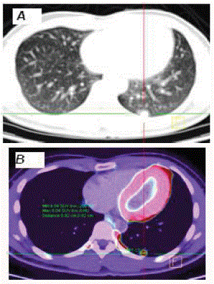
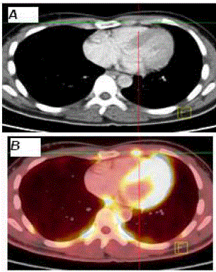
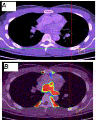
A 17-yr-old man who was diagnosed with par aortic PGL presented with symptoms related to raised serum catecholamine after episodes of tumour recurrences 3 years prior for which patient was treated with surgery. He underwent PET/CT (Biograph Sensation 64, Siemens Medical Systems, Germany) for tumour restaging 1 hr after injection of 10 mCi, Fluorine-18, flurodeoxyglucose- (18F-FDG). 18F-FDG PET/CT demonstrated multiple FDG-avid lesions in the left lung, along the posterior parietal pleural in the left lower hemi thorax, and in the left anterior chest wall (Figure1). Interestingly, all the FDG-avid nodules except a nodule in the lower lobe of the left lung are false positive lesions as evaluated on the correlated CT images (Hounsfield unit: -10) (Figure 2,3). The false FDG-positive lesions are in keeping with FDG-avid brown fat uptake. This patient was referred for further management of metastatic malignant PGL in the lung.
Diagnosis and Discussion
We document multiple foci of FDG brown fat uptake in rare isolation of the anterior chest wall
and parietal pleural which may masquerade malignancy on 18F-FDG PET/CT. The diagnosis was
made on the basis of their fat density nature on the correlated CT averting the treatment intent for
diffuse chest wall metastatic disease. The patient was treated for metastatic PGL recurrence in the
lung.
Oncologic PET is typically performed with the radiopharmaceutical 18F-FDG (Fluorine -18,
flurodeoxyglucose, a glucose analog which form a favoured substance used to signal the in-vivo
mechanism of increased glucose metabolism by malignant cells. Nevertheless, physiological FDG
uptake in adipose tissue, muscle, and myocardium are also important tenet in cancer imaging
particularly in patient with increased sympathetic activity i.e. paraganglioma [1].
Our finding of hyper metabolic brown fat in the mediastinum occurring in mediastinum may
pose confusion to inexperience PET-CT reader given their rare pattern and encounter in this
region [2]. The usual pattern of brown adipose tissue is seen along the
spinal cord as a par vertebral depot, in the mediastinum, particularly
in the para-aortic area, around the heart, particularly the apex and
infradiaphragmatic depots, particularly in the per renal area [3-5].
Knowledge of this potential pitfall of FDG PET/CT is essential to
avoid mistaking it for progression of chest wall metastases in the
thorax thus help improve the diagnostic interpretation and disease
staging.
Figure 1
Figure 1
Axial CT (Figure 1a) and fused FDG PET-CT in the left (Figure 1b) images of the case described. Hairline marker indicate hypermetabolic metastatic paraganglioma nodule in the lower lobe of the left lung (SUVmax: 8.04).
Figure 2
Figure 2
Axial CT (Figure 1a) and fused FDG PET-CT in the left (Figure 1b)
images of the case described. Hairline markers indicate focus of FDG-avid
brown fat uptake in the left anterior chest wall without perceptible abnormality
on CT.
Figure 3
Figure 3
Axial CT (Figure 1a) and fused FDG PET-CT in the left (Figure 1b)
images of the case described. Hairline markers demonstrate another focal
FDG-avid configuration along the parietal pleura of the left lower hemithorax
without discernible abnormality on CT favouring a brown fat signal.
Acknowledgment
This work was supported by the Research University Grant Scheme (RUGS), University Putra Malaysia (UPM). The authors have declared no conflicts of interest.
References
- Hadi M, Chen CC, Whatley M, Pacak K, Carrasquillo JA. Brown fat imaging with (18)F-6-fluorodopamine PET/CT, (18)F-FDG PET/CT, and (123)I-MIBG SPECT: a study of patients being evaluated for pheochromocytoma. J Nucl Med. 2007;48:1077-1083.
- Yeung HW, Grewal RK, Gonen M, Schoder H, Larson SM. Patterns of [18]F-FDG uptake in adipose tissue and muscle: a potential source of false-positives for PET. J Nucl Med. 2003;44:1789-1796.
- Truong MT, Erasmus JJ, Munden RF, Marom EM, Sabloff BS, Gladish GW, et al. Focal FDG uptake in mediastinal brown fat mimicking malignancy: a potential pitfall resolved on PET/CT. AJR Am J Roentgenol. 2004;183:1127-1132.
- K, Tsubone T, Fujiwara K, Masaki T, Kakuma T, Kang M, Tanaka K, et al. Evaluation of human brown adipose tissue using positron emission tomography, computerised tomography and histochemical studies in association with body mass index, visceral fat accumulation and insulin resistance (Abstract). Obes. 2006; 7:87.
- Bar-Shalom R, Gaitini D, Keidar Z, Israel O. Non-malignant FDG uptake in infradiaphragmatic adipose tissue: a new site of physiological tracer biodistribution characterised by PET/CT. Eur J Nucl Med Mol Imaging. 2004;31:1105-1113.


