Review Article
Clinically Significant Incidental Findings on the Unenhanced CT Portion of PET/CT Studies: the Indian Experience
Maria M D’souza*, Rajnish Sharma, Abhinav Jaimini, Madhavi Tripathi, Anupam Mondal and RP Tripathi
Department of Nuclear Medicine and Allied Sciences, Division of PET Imaging, Institute of Nuclear Medicine and Allied Sciences, India
*Corresponding author: Maria M D’souza, Department of Nuclear Medicine and Allied Sciences, Division of PET Imaging, Institute of Nuclear Medicine and Allied Sciences, India
Published: 09 Oct, 2017
Cite this article as: D’souza MM, Sharma R, Jaimini A,
Tripathi M, Mondal A, Tripathi RP.
Clinically Significant Incidental Findings
on the Unenhanced CT Portion of PET/
CT Studies: the Indian Experience. Clin
Oncol. 2017; 2: 1354.
Abstract
Background: The unenhanced CT part of a PET/CT examination is undoubtedly useful for
anatomical localization and attenuation correction. Our study was undertaken with the objective
of evaluating the incidental findings from the unenhanced CT portion of PET/CT studies through
independent interpretation by an expert CT radiologist and to assess their clinical significance.
Methods: Standard PET/CT image acquisition protocol was used on a total of 300 patients. The
unenhanced CT studies were read without prior knowledge of findings from PET and PET/CT
fused images, independently by an experienced radiologist. All recorded findings were classified as
being of major, moderate, or minor significance, corresponding to definitions previously used in
similar studies.
Results: Unenhanced CT revealed potentially clinically significant incidental findings in 11 patients,
findings of moderate clinical importance in 15 patients and those of minor clinical importance in
214 patients. The first group largely included patients with neoplastic processes (both metastatic
and synchronous primary) which were not detected solely on the PET images. There was a high
proportion of tuberculosis (both active and healed) in the latter two groups.
Conclusions: Incidental CT findings are commonly encountered in the course of a PET/CT
examination. A small but significant proportion of cases have findings of major clinical significance,
which may be detected only based on careful analysis of the CT component. Also, there is a high
incidence of tuberculosis (both active and healed) in the Indian setting, which would have important
clinical implications. Thus, skilled interpretation of the complete evaluation.
Keywords: Incidental findings; Unenhanced CT; PET/CT
Introduction
PET scan, or Positron Emission Tomography, is a powerful tool which has made a significant
impact in the field of oncology, cardiology, neurology and imaging of inflammatory disorders. To
put it very simply, it has the ability to detect sites of high metabolic activity. Since many cancers
have significantly higher metabolism than normal tissues or noncancerous masses, PET allows
sensitive detection of even small cancers. 18F-FDG PET enables diagnosis, staging, and restaging of
many cancers with accuracies ranging from 80% to 90% and is often more accurate than anatomic
imaging [1]. FDG PET is a strictly functional modality and lacks anatomic landmarks for precise
morphologic orientation. However, the lack of anatomic landmarks and limited spatial resolution
of PET can make precise anatomic localization challenging, thus limiting accurate evaluation of
cancer patients.
PET-CT Fusion is a newer refinement of the technique that allows the most accurate correlation
of anatomic information (from the CT) and metabolic information (from the PET scan).A
dual-purpose imaging device, PET/CT is literally the combination of PET (positron emission
tomography) and CT (computed tomography) imaging techniques within a single machine. The
individual scans, which are taken virtually simultaneously, can be presented separately or as a single,
overlapping, or "fused" image. This fused image provides a more reliable and much more effective
alternative to the traditional side-by-side visual comparison of PET and CT images. The use of PET/
CT has been advocated as a first-line imaging modality for whole-body tumor staging and restaging
and for assessing response to therapy in different types of cancer [2].
However, although rapid attenuation correction and precise
anatomic localization are clearly useful, the value of the independent
CT interpretation is not clear, especially because many CT scans
obtained as part of PET/CT are obtained at somewhat lower power
settings than are standard CT scans and with no or limited use of
contrast material. Currently, the CT portion of PET/CT in many
centers is a lowered-dose, unenhanced study and used only for image
fusion and attenuation correction. Potentially useful information
can be added through independent readings of the unenhanced-CT
portion of PET/CT by a radiologist skilled in CT. Our study was
undertaken with the objective of evaluating the incidental findings
from the unenhanced CT portion of PET/CT studies through
independent interpretation by an expert CT radiologist and to assess
their clinical significance.
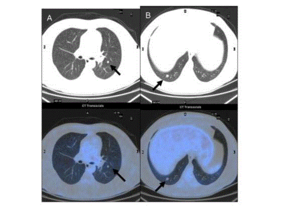
Figure 1
Figure 1
Follow-up case of colon carcinoma showing subpleural nodules on
CT (upper row) in left (A) and right (B) lung fields, with no evidence of FDG
uptake on the fused PET/CT image (lower row).
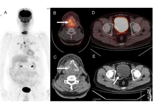
Figure 2
Figure 2
A case of laryngeal carcinoma. MIP image (A) shows increased
uptake at the primary site with intrathoracic spread. The FDG avid
invasive laryngeal mass is well seen on the PET/ CT and CT image (B,C).
Additionally, an unsuspected bladder mass is seen on the CT image (E),
which gets obscured on the PET/CT image by the physiological high uptake
in the bladder (D).
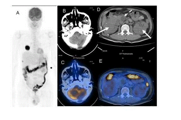
Figure 3
Figure 3
A follow-up case of cerebellar hemangioblastoma. MIP image (A)
shows FDG avid metastatic foci in the lung and liver. Axial CT (B) shows
a post-operative defect in the left cerebellar hemisphere with reduced
adiotracer uptake on the PET/CT image (C). Unsuspected bilateral renal
masses (arrows) and pancreatic cysts (dotted arrows ) seen on CT (D), none
of which show increased radiotracer activity on the PET/CT fusion image
(E). This unusual constellation of findings points towards the rare von Hippel
Lindau syndrome.
Methods
The study included a total of 300 sequential patients (139 men
and 161 women; mean age, 52.3 y) referred for clinical evaluation
of known or suspected cancer. The institutional review board
approved of this prospective study. All subjects fasted for six hours
prior to the study. They were administered 10mCi (370 mBq) 18FFDG
intravenously followed by a period of rest of 60 minutes with
minimal body activity. The PET/CT scans were performed using a
whole body Full Ring PET camera (Discovery STE16-GE). Wholebody
CT scanning was performed by a continuous spiral technique
using a 16-slice helical CT with a gantry rotation speed of 0.8 second.
No intravenous or oral contrast agent was used. After the CT scan, an
emission scan was performed from the head to thigh for 2 minutes per
frame. The attenuation-corrected PET images using the CT data were
reconstructed by using an ordered subset expectation maximization
algorithm (20 subsets, 4 iterations),and they were displayed in a 128
x 128 matrix. Accurate co registration of the separate CT and PET
scan data was performed. Images were evaluated by an experienced
nuclear medicine physician.
The unenhanced CT studies were read on an ADW (Advantage
Window) workstation without prior knowledge of findings from
PET and PET/CT fused images, independently by an experienced
radiologist. Details were recorded concerning the nature of the
abnormality, the anatomic location, and any information from
previous and subsequent imaging studies. Findings from unenhanced
CT were considered significant if they were not detected or explained
by PET findings and were considered to clearly require additional
work-up. All recorded findings were classified as being of major,
moderate, or minor significance, corresponding to definitions
previously used in similar studies [3,4]. A finding was considered of
major importance when the CT appearance was highly suspicious for
an abnormality relevant to the immediate treatment of the patient.
Abnormalities were considered of moderate importance when the
imaging appearances suggested clinically important disease but
required further correlation with other clinical or imaging findings
and required eventual medical or surgical treatment. Abnormalities
were considered of minor importance when the imaging appearances
did not suggest clinically important disease and did not require
additional workup.
Results
Unenhanced CT revealed potentially clinically significant
incidental findings in 11 patients. Sclerotic metastases were noted in
four cases, multiple small subpleural pulmonary metastases in three
(Figure 1), cerebral metastasis in one, synchronous malignancy in
the bladder (Figure 2) and the kidney in one each. Additionally, one
patient had a spectrum of findings pointing toward a rare syndrome
– namely von Hippel Lindau syndrome (Figure 3). None of these
findings were detected on PET scans viewed in isolation.
Findings of moderate clinical importance were noted in 15
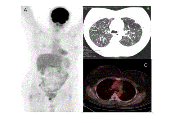
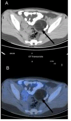
patients which included pulmonary tuberculosis in 9 patients (Figure
4), calcified left ventricular aneurysm, adrenal adenoma, dermoid
cyst (Figure 5), renal Angiomyolipoma, misplaced intrauterine
contraceptive device and tubercular cervical lymphadenopathy in
one patient each.
There were findings of minor clinical importance in 214 patients
which included abnormalities such as gallstones, renal stones,
hiatus hernia, abdominal hernia, renal cysts, healed pulmonary
Kochs (Figure 6), calcified granulomas in the brain, uterine fibroids,
hepatic cysts, prostate enlargement, spondylolisthesis, spondylosis,
bone islands, solitary bone cysts, hemangiomas and old posttraumatic
changes in bones. Some of these features had already been
documented based on previous investigations and did not warrant
alteration in management. However, it is important to document
these abnormalities as they may explain the symptoms which the
patient may develop in the course of follow-up, which is unrelated to
the present disease.
Figure 4
Figure 4
A follow-up case of choriocarcinoma. No residual disease seen
on MIP image (A). However CT image (B) shows evidence of military
tuberculosis which shows no radiotracer uptake on fused PET/CT image (C).
Figure 5
Figure 5
A follow-up case of carcinoma breast. CT image (A) shows a
rounded well-defined lesion of fat attenuation suggestive of dermoid cyst. No
radiotracer uptake noted on the PET/CT fusion image (B).
Figure 6
Figure 6
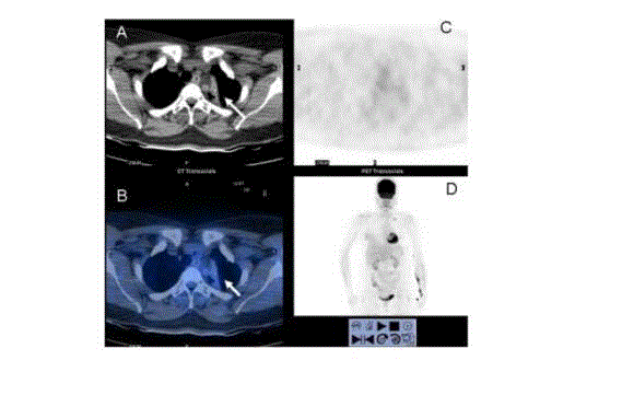
A follow-up case of carcinoma breast. A fibro-calcific lesion is seen
in the upper lobe of left lung seen on CT (A). No radiotracer uptake noted on
the PET and fused PET/CT images (B, C). MIP image (D) shows evidence of
left mastectomy with left supraclavicular lymph-node.
Discussion
Clinically significant findings from the unenhanced CT portion
of PET/CT are relatively infrequent (3%) but could be serious enough
to warrant major alterations in clinical management [5]. Thus, we
believe it is most appropriate for the CT portion to be interpreted by
a physician skilled in CT interpretation with special attention to the
lesions that PET alone can fail to detect.
The importance of potential findings on the CT component of
PET/CT is reflected in a recent paper from the American College
of Radiology [6] regarding interpretation of PET/CT studies.
It is recommended that the interpreting radiologist or nuclear
physician report additional findings present on CT images, even if
the examination is performed purely for anatomic localization only.
There is much debate about who should interpret the studies, but it is
clear that the CT component of the study cannot be ignored.
Most of the additional findings detected in our study were
classified as being of minor importance, which are extremely common
in the general population and do not warrant additional investigation
or follow-up. However a noteworthy feature is the large proportion
of patients who had features of healed tuberculosis, be it in the lungs,
the mediastinum or the brain. This is a finding that is closely related
to the high prevalence of tuberculosis in our Indian population. What
is worrisome is, that many of these patients are at a risk of developing
reactivation owing to the immunosuppression which occurs as an
inevitable part of their chemotherapeutic regimens. The relatively
large number of patients with active tuberculosis in our study group
lends credence to this. Early diagnosis and prompt institution of
treatment would lead to early amelioration of symptoms of this
completely curable disease. Acute or chronic inflammation, abscesses,
inflammatory lymphadenopathy and non-specific reactions following
radiotherapy may mimic tumor tissue in PET scans [6-8]. However,
analyzing the pattern of disease on the CT component enables
distinguishing inflammatory from neoplastic process in the majority
of cases. This holds specially true for tuberculosis.
The other important feature is the detection of synchronous
malignancy and of distant metastasis which fail to get picked up on
PET/CT. The lack of FDG avidity at these sites could be due to several
reasons - small size as in the case of pulmonary metastasis, location
as in the case of cerebral metastasis and bladder malignancy, and
low affinity for the radiopharmaceutical as in the case of mucinous
metastasis. It is already known that PET has relatively low sensitivity
for cerebral metastatic lesions, mainly because of the limited spatial
resolution of PET scanners and the intense metabolic activity already
present throughout the rest of the cortex [9,10]. In certain cases, brain
metastases appear as areas of reduced 18F-FDG uptake compared
with the surrounding cortex [11]. In addition, it should be recognized
that certain tumors with variable 18F-FDG uptake, such as prostate
cancer, hepatocellular carcinoma, renal cell carcinoma, bladder
carcinoma and osteoblastic bone metastases [12-14], may be more
readily detected owing to their abnormal appearance on CT images
than owing to their PET appearance.
Conclusions
Incidental CT findings are commonly encountered in the course of a PET/CT examination. Although this may not have a direct bearing on the immediate management in every case, it would have an impact on the overall clinical profile of the patient. A small but significant proportion of cases have findings of major clinical significance, which may be detected only based on careful analysis of the CT component. Also, there is a high incidence of tuberculosis (both active and healed) in the Indian setting, which would have important clinical implications. Thus, skilled interpretation of the unenhanced CT component of the PET/CT scan is a vital pre-requisite to complete evaluation.
References
- Bar-Shalom R, Yefremov N, Guralnik L, Gaitini D, Frenkel A, Kuten A, et al. Clinical performance of PET/CT in evaluation of cancer: additional value for diagnostic imaging and patient management. J Nucl Med. 2003;44:1200-1209
- Pfister DG, Johnson DH, Azzoli CG. American Society of Clinical Oncology treatment of unresectable non-small cell lung cancer guideline. J Clin Oncol. 2004;22:330 -353.
- Hara AK, Johnson CD, MacCarty RL, Welch TJ. Incidental extracolonic findings at CT colonography. Radiology.2000; 215: 353 -357.
- Hellstrom M, Svensson MH, Lasson A. Extracolonic and incidental findings on CT colonography (virtual colonoscopy). AJR. 2004;182:631-638.
- Osman MM, Cohade C, Fishman EK, Wahl RL. Clinically Significant Incidental Findings on the Unenhanced CT Portion of PET/CT Studies: Frequency in 250 Patients. J Nucl Med. 2005; 46: 1352-55.
- Coleman RE, Delbeke D, Guiberteau MJ. Concurrent PET/CT with an integrated imaging system: intersociety dialogue from the Joint Working Group of the American College of Radiology, the Society of Nuclear Medicine, and the Society of Computed Body Tomography and Magnetic Resonance. J Nucl Med. 2005;46 : 1225-1239.
- Valk P, Pounds T, Tesar RD. Cost effectiveness of PET Imaging in Oncology. Nucl Med Biol.1996; 23:737-43.
- Strauss LG. Sensitivity and specificity of positron emission tomography (PET) for the diagnosis of lymph node metastasis. Recent Results Cancer Res. 2000;157:12-9.
- Bakheet SM, Powe J, Ezzat A. F-18-FDG uptake in tuberculosis. Clin Nucl Med. 1998; 23 (11): 739-42.
- Coleman RE. PET in lung cancer staging. Q J Nucl Med. 2001 45:231-234.
- Rohren EM, Provenzale JM, Barboriak DP, Coleman RE. Screening for cerebral metastases with FDG PET in patients undergoing whole-body staging of non-central nervous system malignancy. Radiology. 2003; 226:181 -187.
- An YS, Yoon JK, Lee MH, Joh CW, Yoon SN. False negative F-18 FDG PET/CT in nonsmall cell lung cancer bone metastases. Clin Nucl Med. 2005; 30:203 -204.
- Kang DE, White RL Jr, Zuger JH, Sasser HC, Teigland CM. Clinical use of fluorodeoxyglucose F 18 positron emission tomography for detection of renal cell carcinoma. J Urol 2004;171 : 1806-1809.
- Liu IJ, Zafar MB, Lai YH, Segall GM, Terris MK. Fluorodeoxyglucose positron emission tomography studies in diagnosis and staging of clinically organconfined prostate cancer. Urology. 2001;57:108 -111.

