Case Report
A Case of Invasive Adenocarcinoma Arising from Heterotopic Pancreas in the Stomach and Review of the Literature
Roberto Bini1, Franco Coppola3, Antonella La Terra3, Tiziana Viora1, Renzo Leli1 and Alfredo Addeo2*
1Department of General and Emergency Surgery, SG Bosco Hospital, Italy
2Department of Oncology, Bristol Cancer Center, UK
3Department of Gastrointestinal Endoscopy, SG Bosco Hospital, Italy
*Corresponding author: Alfredo Addeo, Department of Oncology, Bristol Cancer Center, UK
Published: 14 Sep, 2017
Cite this article as: Bini R, Coppola F, La Terra A, Viora
T, Leli R, Addeo A. A Case of Invasive
Adenocarcinoma Arising from
Heterotopic Pancreas in the Stomach
and Review of the Literature. Clin
Oncol. 2017; 2: 1348.
Abstract
Heterotopic pancreas is a rare condition of tissue found outside the pancreas without anatomic connection with ectopic one. Generally asymptomatic and incidentally findings could be located elsewhere in the gastrointestinal tract. Degeneration into neoplasm is rare. Less than sixteen cases has been describe. Our case is the first well reported an adenocarcinoma arising from a locally advanced stomach’s HP discovered for hematemesis.
Introduction
The Heterotopic Pancreas (HP) is defined as a pancreatic tissue found outside the eutopic
pancreas without anatomic or vascular connections between them.
It is not uncommon with an incidence of approximately 0.5% - 13.7% in autopsy studies and
0.5% in upper abdominal laparotomies and It can be found anywhere in the gastrointestinal tract.
The most common site is the stomach, mostly in the antrum and prepyloric region on the greater
curvature or posterior wall. The heterotopic pancreas is usually an incidental finding, however,
mass-like manifestations causing pyloric obstruction, ulcer, and bleeding can be observed in the
gastric area [1].
We report a case of a patient with HP of the stomach who developed an invasive adenocarcinoma
presenting with hematemesis.
Case Report
A 76 years old woman, with a past medical history of hypertension, under evaluation for
anemia, was admitted to the Emergency Department (ED) with melena and hematemesis.
Hemoglobin was 6.2 g/dl, red blood cell 2.3 x 10⁶, Hematocrit 22%, elevated lactate and Base Deficit
(BD). Her blood pressure was 75 over 55 mmHg and 120 heart pulse rate, euthermic, skin pale and
clammy. Coagulation screening was within normal value. She received fluids and blood units. She
subsequently underwent Oesophago-Gastro-Duodenoscopy (OGD) that revealed the presence of
a blood clot occupying the body of the stomach. During the admission she became unstable so an
angio-Computed Tomography (CT) was performed. That showed a neoplastic lesion involving the
body of the stomach, transverse colon, distal pancreas and spleen (Figure 1). Due to presence of
multiple collateral vessels feeding the lesion with high risk of massive necrosis embolization was not
performed. A gastrectomy, spleno-pancreatectomy and resection of the transverse colon with Roux
and Y esophagojejunostomy and end to end colo-colic anastomosis was perfomed.
In the first post-operative period the patient developed Acute Respiratory Distress Syndrome
(ARDS) and required external ventilation and hemodynamic support in intensive care unit. The
histology revealed/showed adenocarcinoma (pTN4N2M0) (Figure 2 and 3). After 24 days the patient
was discharged and referred to the oncological department. Due to comorbidity she wasn’t deemed
fit for any oncological therapy and, after one year, she died for the progression of her disease.
Discussion
HP is often and incidental finding and many patients with HP are asymptomatic and require
no treatment [2]. The rate of HP during autopsy varies from 0.6% to
15% and about 75% of ectopic pancreases are found in the stomach,
duodenum, or ileum [3]. In the stomach, HP is most often located
along the greater curve of the prepyloric antrum and especially within
6 cm of the pyloric ring. HP are usually less than 4 cm in size, and
a central mucosal depression is often recognized at endoscopy. An
ectopic pancreas usually invades the submucosal or muscle layer, and
has histologically normal pancreatic tissue [4].
Histology confirmation and diagnosis that a malignant tumor
has raised from pre-existing ectopic pancreatic tissue may be rather
difficult. Described three criteria to prove that a malignancy arose
from an ectopic pancreas: 1) the tumor must be within or near the
ectopic pancreatic tissue; 2) a direct transition between pancreatic
structures and carcinoma must be observed; 3) the non-neoplastic
pancreatic tissue must, at a minimum, be fully developed [5].
Heinrich classified the heterotopic pancreas into 3 types [6]:
type I, all the components of the pancreas including ducts, acini, and
endocrine islets; type II, ducts with acini; and type III, ducts with a
few acini or dilated ducts only, so called adenomyoma. When the
pancreaticobiliary-type ducts predominate, they are often surrounded
by hypertrophic smooth muscle bundles.
In a series of 32 cases ofsymptomatic HP none of the patients
was diagnosedpreoperatively [7]. Barium swallow studies may
show arounded filling defect, sometimes with central indentation.
Imaging studies with endoscopic ultrasound (EUS) and (CT) are
frequently used for thediagnosis of gastrointestinal submucosal
tumor and can behelpful in the diagnosis of gastric HP, but are not
specific [8]. However, studies have suggestedthat both EUS and CT
help to distinguish HP from othersubmucosal tumors. Endoscopic
appearance of HP is thatof a well circumscribed submucosal mass
with a normaloverlying mucosa and a central dimpling which
correspondswith the opening of a duct. The characteristic dimplingor
umbilication is observed in less than half of the cases,and therefore,
HP may easily be misinterpreted as anothersubmucosal tumor such
as stromal tumor, or leiomyoma atendoscopic examination. Because
GISTs are by far the mostcommon gastric submucosal tumors, HP can
frequently bemistaken for GIST at endoscopy as happened in our case
[9]. Endoscopic biopsy performed by using standardbiopsy forceps
is most often unremarkable. However, a few reported cases of HP
were diagnosed with biopsies obtained with the jumbo forceps [10].
Diagnosis can occasionally be made through endoscopical biopsies
but those are inconclusive in about 50% of the cases as normal gastric
mucosa could cover the HP [11].
EUS-guided aspiration has been reported to be helpful to
diagnose HP [12]. If an endoscopic resection is considered, EUS is
also extremely useful for pre-excision assessment. There was no
correlation between the histological type of HP and the presence
of symptoms. Surgery is frequently needed to make a definitive
diagnosis and to plan further treatment because the differential
diagnosis includes leiomyoma, lymphoma, carcinoid tumors, and
other malignancies.
If HP is diagnosed as an incidental finding, local excision is
recommended. Symptomatic HP should be resected. Endoscopic
excision can be considered in select cases depending on the size and
location of the mass, especially for treating the benign lesions of HP.
Because the preoperative imaging studies (ultrasonography,
endoscopic ultrasonography and computerized tomography) are
not very specific [13], intraoperative frozen section studies are
useful to confirm the diagnosis and exclude the most common
differential diagnosis of HP such as gastrointestinal stromal tumor,
gastrointestinal autonomic nerve tumor, lymphoma, carcinoid
tumor and other malignancies. Recently, the combination of
endoscopic ultrasonography with fine needle aspiration cytology
from submucosal lesions is showing encouraging results due to its
sensitivity which ranges from 80% to100% [14].
The histogenesis of HP tissue is controversial. Yamagiwa et al.
[15] suggested that Heinrich type I HP of the stomach was caused
by migration from fetal pancreas, while Heinrich type II and III
heterotopic pancreas arises through erroneous differentiation of
primitive gastric mucosal epithelium. The real incidence of HP into
the stomach is uncertain; in a large series [15,16]. They examined the
whole stomach by numerous sections in gastrectomies. They showed
107 cases of heterotopic pancreas out of 5,446 surgically resected
stomachs; the incidence was 1.2%. Reported 12.7% incidence of
cancer in their series of HP in the 1980 [17].
The review of the literature, showed 14 cases well documented
of neoplasm arising from pancreatic ectopia in the stomach (Table
1). The site most involved is the antrum and generally the lesion was
well confinated into in stomach wall. Surgery ranged from small
resection to total gastrectomy. Overall survival was good. No case of
diffuse cancer as our case has been described in the past. The majority
of carcinomas arising with in HP are adenocarcinoma sora plastic
carcinomas. Review of the literature reveals adenocarcinomas arising
within heterotopic pancreas to have a better prognosis than patients
with adenocarcinoma of the pancreas.
In conclusion, based on the literature the findings of HP is not so
uncommon. The gastric localization of HP is most common among
the number of HP, but, based on literature’s data, more than ten
cases developed cancer. The correct approach for diagnosis includes
endoscopic ultrasound with several biopsies, fine needle aspiration
and CT scan. A resection remains the gold standard if HP is suspected.
Figure 1
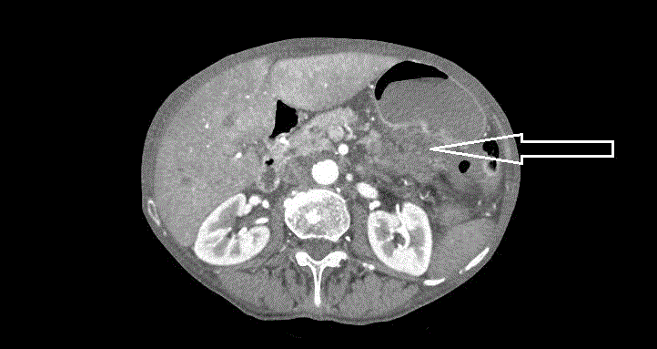
Figure 1
CT scan of the neoplasm arising from EP in stomach (white arrow).
Figure 2
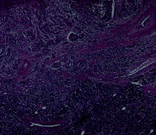
Figure 2
Haematoxylin and Eosin 100X. Ectopic pancreas.
Figure 3
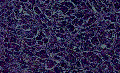
Figure 3
Haematoxylin and Eosin 50X. Passage from HP (in the lower
portion of the image) and neoplasm (in the upper part of the image).
Table 1

Table 1
Lists of article concerning Neoplasm arising from HP in the stomach.
References
- Tanaka K, Tsunoda T, Eto T. Diagnosis and management of heterotopic pancreas. Int Surg. 1993; 78(2): 32-35.
- Lai EC, Tompskins RK. Heterotopic pancreas: review of a 26 year experience. Am J Surg. 1986; 151(6): 697-700.
- Hickman DM, Frey CF, Carson JW. Adenocarcinoma arising ingastric heterotopic pancreas. West J Med. 1981; 135(6): 57e62.
- Jeong HY, Yang HW, Seo SW, Seong JK, Na BK, Lee BS, et al. Adenocarcinoma Arising from an Ectopic Pancreas in the Stomach. Endoscopy. 2002; 34(12): 1014-1017.
- Guillou L, Nordback P, Gerber C. Ductal adenocarcinoma arising in a heterotopic pancreas situated in a hiatal hernia. Arch Pathol Lab Med. 1994; 118(5): 568.
- von Heinrich H. Ein Beitrag zur Histologie des sogen: Akzessorischen Pankreas. Virchows Arch A Pathol Anat Histopathol. 1909; 198: 392-401.
- Pang LC. Pancreatic heterotopia: a reappraisal and clinicopathologic analysis of 32 cases. South Med J. 1988; 81(10): 1264-1275.
- Cho JS, Shin KS, Kwon ST, Kim JW, Song CJ, Noh SM, et al. Heterotopic pancreas in the stomach: CT findings. Radiology. 2000; 217(1): 139-144.
- Kim JY, Lee JM, Kim KW, Park HS, Choi JY, Kim SH, et al. Ectopic pancreas: CT findings with emphasis on differentiation from small gastrointestinal stromal tumorand leiomyoma. Radiology. 2009; 252(9): 92-100.
- Shaib YH, Rabaa E, Feddersen RM, Jamal MM, Qaseem T. Gastricoutlet obstruction secondary to heterotopic pancreas in the antrum:case report and review. Gastrointest Endosc. 2001; 54(4): 527-530.
- Jiang LX, Xu J, Wang XW, Zhou FR, Gao W, Yu GH, et al. Gastric outlet obstruction caused by heterotopic pancreas:A case report and a quick review. World J Gastroenterol. 2008; 14(43): 6757-9.
- Rodriguez FJ, Abraham SC, Allen MS. Fine-needle aspiration cytology findings from a case of pancreatic heterotopia at the gastroesophageal junction. Diagn Cytopathol. 2004; 31(3): 175-179.
- Gurocak B, Gokturk HS, Kayacetin S. A rare case of heterotopic pancreas in the stomach which caused closed perforation. Neth J Med. 2009; 67(7): 285-287.
- Christodoulidis G, Zacharoulis D, Barbanis S. Heterotopic pancreas in the stomach: A case report and literature review. World J Gastroenterol. 2007; 13(45): 6098-6100.
- Yamagiwa H, Ishihara A, Sekoguchi T, Matsuzaki O. Heterotopic pancreas in surgically resected stomach. Gastroenterol Jpn.1977; 12: 380-386.
- Yamagiwa H, Onishi N, Nishi M. Heterotopic pancreas of the stomach: Histogenesis and immunohistochemistry. Acta Pathol Jpn 1992; 42(4): 249-254.
- Nakaoa T, Yanoh K, Itoh A. Aberrant pancreas in Japan. In: Review of the literature and report of 12 surgical cases. Med J Osaka Univ. 1980; 30(3-4): 57-63.