Review Article
Introduction to 5-Aminolevulinic Acid-Protoporphyrin IXMediated Radiodynamic Therapy (RDT)
Junko Takahashi1* and Hitoshi Iwahashi2
1Biomedical Research Institute, National Institute of Advanced Industrial Science and Technology, Tsukuba, Ibaraki, Japan
2Faculty of Applied Biological Sciences, Gifu University, Gifu, Japan
*Corresponding author: Junko Takahashi, Biomedical Research Institute, National Institute of Advanced Industrial Science and Technology, Tsukuba, Ibaraki, 1-1-1 Higashi, Tsukuba, Ibaraki 305-8566 Japan
Published: 04 Sep, 2017
Cite this article as: Takahashi J, Iwahashi H. Introduction
to 5-Aminolevulinic Acid-Protoporphyrin
IX-Mediated Radiodynamic Therapy
(RDT). Clin Oncol. 2017; 2: 1330.
Abstract
Photodynamic Therapy (PDT) is a light-based method that uses photo-reactive molecules, such as
protoporphyrin IX (PpIX), to ablate tumors. Recently, PpIX was shown to act as a radio-reactive
molecule by enhancing generation of Reactive Oxygen Species (ROS) upon X-ray irradiation.
This characteristic enables radiodynamic Therapy (RDT), which uses radiation as a physical
stimulus instead of light used in photodynamic therapy. The merit of RDT over PDT is the X-ray’s
penetrability through tissues, which will find many applications for treatment of deep cancers. In
this mini-review, we discuss the potential applications of radiodynamic therapy.
Keywords: Radiotherapy; Radiodynamic therapy (RDT); 5-aminolevulinic acid (5-ALA);
Protoporphyrin IX (PpIX); Photodynamic therapy (PDT)
Introduction
Surgical resection, when feasible and safe, should be performed and in many cancers stands as
the first line treatment. Depending on cancer location, the role of radiotherapy is underestimated but
cannot replace existing therapies. However, acute toxicity and potential long-term adverse effects
often limit the dose of radiation to levels that are insufficient for controlling tumors. Radiosensitizers
have the ability to specifically increase tumor radiosensitivity [1-5], and the radiosensitizing effects
of cis-platinum and other chemotherapeutic agents have been previously reported [6-9]. Although
some clinical benefits have been achieved, the use of these compounds did not significantly improve
outcomes compared to radiation treatment alone.
Photodynamic therapy (PDT) is becoming a more widely accepted modality for treating solid
tumors [10-11]. Topical or systemic administration of a photosensitizer leads to its accumulation
and retention in tumors, and accumulated photosensitizers can be subsequently activated by a
specific wavelength of light. Upon light activation, an excited photosensitizer undergoes type I
(electron transfer) and/or type II (energy transfer) reactions to produce highly Reactive Oxygen
Species (ROS), resulting in apoptosis and/or necrosis of exposed cells [12–13]. The most important
pathways for clinical PDT are the generation of type II photochemical reactions. In these, a
photosensitizer interacts with oxygen to generate 1O2, which is considered to be essential for PDT’s
ability to ablate tumors [11,13].
Since the 1950s, in vivo studies have shown that porphyrins can modify the effects of ionizing
radiation [14,15]. Photofrin and hematoporphyrin derivatives are known to be a mixture of
porphyrins formed by acetic acid-sulfuric acid treatment of hematoporphyrin. In 1981 Dougherty et
al. [16] described a gel filtration procedure to isolate hematoporphyrin derivatives in relatively pure
form. But being insufficiently purified, Photofrin and hematoporphyrin derivatives were chemically
heterogeneous. Although porphyrins, under certain conditions, seemed to be act as radiosensitizers,
the observed radiosensitizing effects were considered not due to the main components but rather to
minor ones [17]. There is some contradicting evidence in the literature, because both protective and
sensitizing effects have been reported for porphyrins [18]. Until recently, the radiosensitizing effects
of the porphyrins have been studied under biological settings, and these studies were inconclusive.
Recently, protoporphyrin IX (PpIX) was characterized as a radio-responsive compound
and shown to produce ROS upon X-ray irradiation [19]. This discovery demonstrated the use of
radiation as a physical stimulus for radiodynamic therapy (RDT), instead of laser light used for
photodynamic therapy (Scheme 1). RDT has already been tested, both in vitro and in vivo, and
it is currently being tested in clinical trials [20]. The merit of RDT
over PDT is the X-ray’s penetrability through tissues, which will find
many applications for treatment of deep cancers. This review aims
to describe the mechanisms which could be assumed as general for
the radiosensitizers, those attributable to porphyrin-type, underlying
RDT.
Scheme 1
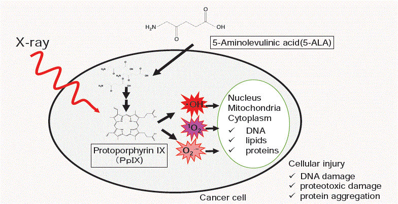
Scheme 1
Schematic illustration of radiodynamic therapy. PpIX contributes to enhanced generation of ∙OH, O2
∙−, and 1O2 in the presence of X-ray irradiation. 1O2 is thought to be a major ROS produced by photodynamic therapy.
PpIX Produces ROS Upon X-ray Irradiation by Physicochemical Reactions
PpIX generates ROS effectively upon X-ray irradiation [19]. ROS generation was monitored using two ROS detection reagents: 2-[6-(4-amino)phenoxy-3H-xanthen-3-on-9-yl] benzoic acid (APF) [21], and dihydroethidium (DHE) [22], with ethanol as a quencher of ·OH in solutions containing different concentrations of PpIX. Upon X-ray irradiation, ·OH and 1O2 were detected by APF, while the generation of O2·− and/or 1O2 was detected by DHE. While the APF reaction was quenched by the addition of ethanol, the DHE reaction was not. Upon UV irradiation, O2·− and/or 1O2 were generated, and these results were consistent with those of previous reports [11]. These results confirmed that X-ray irradiation with PpIX enhanced generation of ·OH, O2·−, and 1O2 for potential use in RDT, as well as UV irradiation with PpIX generated 1O2 and/or O2·− for PDT applications [19]. To study the role of PpIX in mitochondrial metabolism of H2O2, Zeng et al. [23] analyzed catalitic conversion of H2O2 into 1O2 by PpIX. In their experiment, no 1O2 was produced in the presence of only PpIX without X-ray irradiation, but 1O2 was produced with irradiation. The 1O2 formation appeared to increase with an increase in the concentration of H2O2. They might observe the reactions of PpIX and X-ray irradiation unintentionally.
Intracellular ROS Generation by X-ray Irradiation of PpIX
The effects of ROS generated by X-ray irradiation of PpIX were
evaluated in cancer cells in vitro. Intracellular ROS production by PpIX
was assessed by pre-incubating the HeLa cells with cell-permeable
fluorescent probes (APF or DHE) with different sensitivities to specific
ROS species (∙OH, O2·−, and 1O2). ROS levels increased with increasing
X-ray doses and intracellular PpIX concentrations, according to both
APF and DHE detection systems [24]. Gene expression profiling was
performed using DNA microarray. Differences in gene expression
were observed in HeLa cells treated with PpIX or X-rays only, and
cells treated with PpIX and X-ray radiation together. PpIX and X-ray
irradiation treatment together induced systematic changes in the
expression of genes related to cell-cycle arrest and inhibition of DNA
replication. Treatment PpIX and X-ray irradiation together resulted
in higher numbers of genes with altered expression profiles relative
to control than treatment with X-ray irradiation only; however, the
qualitative gene expression remained the same between the two
conditions. These results suggest a complex mechanism by which
PpIX enhances ROS generation during application of X-rays. As a
result, cell viability decreased by inducing DNA damage and cellcycle
arrest.
Yamamoto et al. [25] showed that intracellular PpIX induced
by 5-aminolevulinic acid (5-ALA) plays an important role in
radiosensitization upon ionizing irradiation. Intracellular ROS
generated by ionizing radiation or heme content in glioma 9L cells
was detected by dichlorofluorescein (DCF) fluorescence using the
oxidant-sensitive probe 2’,7’-dichlorofluorescin diacetate (DCFD).
Oxidation of DCFH is known to be not always related the generation
of ROS but may be related heme content in cells [26,27]. Pretreating
cells with 5-ALA before irradiation resulted in increased DCF
fluorescence. Interestingly, the DCF fluorescence generated by
ionizing irradiation predominantly coincided with 5-ALA-induced
PpIX accumulation in the cytoplasm of 9L cells [25]. Intracellular
ROS levels were significantly higher in 9L cells 12 h after applying
ionizing irradiation than in those measured immediately after
applying ionizing irradiation, and 5-ALA pretreatment strongly
enhanced ROS levels [28].
PpIX Enhances Cancer Radiotherapy in a Tumor Model
To evaluate the tumor-suppressive effects of PpIX as a radiosensitizer, a B16-BL6 tumor model was established in C57BL/6 J mice [29]. Porphyrin accumulation in implanted B16-BL6 tumors 24 h after at a topical dose of 50 mg/kg 5-ALA administration was 6.2 times higher than with systematic, local administration providing efficiencies equal or surpassing those of PDT (3.0 ± 1.4 μg per gram wet weight). Using a protocol in which ALA was administered immediately after X-ray irradiation for convenient preparation for the next irradiation, tumor suppression significantly improved in animals treated with 5-ALA and fractionated doses of X-ray irradiation (3Gy × 10; total, 30 Gy). Microarray analyses of tumor tissues collected after 10 sessions of fractionated irradiation co-treated with or without 5-ALA revealed that the majority of dysregulated genes were related to cell-cycle arrest. Co-treatment with 5-ALA affected the level of expression but not the pattern of gene expression [30]. These results suggest that irradiation treatment leads to PpIX-mediated ROS generation. Yamamoto et al. [31] also showed that 5-ALA-induced generation of PpIX upon fractionated X-ray dosing (2 Gy × 5; total, 10 Gy) enhances the antitumor response and strongly inhibits growth of 9L gliomas in rats.
Other Mechanisms Underlying Radiosensitization by Porphyrins
Lemay et al. showed substituting cationic, N-propyl porphyrins
with bromines onto the propyl side-chains substantially enhances
the radiosensitizing potency of the parent compound. As a similar
radiosensitizing effect was detected for different radiation energies,
they thought that the high energy photons could be used to treat
tumors in conjunction with the radiosensitizer [32].
Porfimer sodium, sold as Photofrin, and hematoporphyrin
are used as photosensitizers in PDT, and also thought to be
radiosensitizers [17,33-36]. Schaffer et al. [33] showed that Photofrin
significantly improved the tumor responses to 5 Gy and 15 Gy
radiation doses in mice implanted subcutaneously with human
bladder cancer RT4 cells. Two cell lines, RT4 and U-373 MG, treated
with Photofrin before radiation had lower survival than untreated
cells, which were treated with irradiation only under identical
conditions [34]. Luksiene et al. [17] examined the radiosensitizing
properties of three porphyrin-type compounds: hematoporphyrin
dimethyl ether, Photofrin, and hematoporphyrin derivatives,
and found that hematoporphyrin dimethyl ether was the most
effective of the three (hematoporphyrin dimethyl ether > Photofrin
> hematoporphyrin derivatives). The authors showed that only
aggressive Ehrlich Ascites Carcinomas (EAC) were radiosensitized
by porphyrins, and ligands of peripheral benzodiazepine receptors
(PBR) may diminish cell growth in aggressive EAC. They thought
that porphyrins, being ligands of PBR, which are highly expressed in
aggressive tumors only, can inhibit tumor cell proliferation and act in
concert with ionizing radiation [17]. Benayoun et al. [35] showed that
Photofrin treatment radiosensitizes the tumor-initiating U-87MG
cells derived from human glioblastoma and improves outcomes
when combined with radiotherapy in vitro and in vivo. However, the
corresponding mechanisms of action of Photofrin or its derivatives
remain unknown. Lam et al. [36] found that PDT treatment before
external-beam radiotherapy improved survival in a randomized
comparative analysis of the safety and efficacy of PDT, using
Photofrin combined with palliative radiotherapy in patients with
obstructive endobronchial tumors. In this case, Photofrin was used
as a photosensitizer, not as a radiosensitizer, trial studies combining
photosensitizers and radiotherapy in patients are underway. If X-ray
irradiation were done during photosensitizer accumulated in tumor,
it might act as radiosnsitizer unintentionally.
In the aspect of radiosensitizing effects to cells, the complexity of
the radiosensitizing activity of PpIX to the essential biological factors
must be considered including conditions occurring in each systems.
Sailer et al. [37] showed that the different PDT efficacy of 5-ALA
induced PpIX was related to the different intercellular location of
glioblastoma, breast cancer and ovarian cancer cells. It is known that
the cytotoxic ROS, singlet oxygen or superoxide radicals, react with
various biomolecules (e.g. proteins) and caused cell damages in PDT.
Maitra et al. [38] showed that external and internal porphyrinogenic
stress caused proteotoxic damage and protein aggregation. On the
other hand, X-ray induced ∙OH caused DNA degradation, and PpIX
enhance generation of ∙OH in addition to 1O2, O2·−, cytotoxic effect of
RDT might be DNA and protein damage.
The modification of spectral properties of PpIX were monitored
as the specific indicators of the PDT effect [39,40]. Dysart and
Patterson showed that fluorescence photobleaching of ALA induced
PpIX exhibited complex photobleaching kinetics in vitro, likely
resulting from differential binding of the sensitizer within the cell
[39]. In RDT, the changes in absorption spectrum and fluorescence
emission spectrum of PpIX were observed with the increase of X-ray
irradiation dose [30].
The essential questions concerning to the enhanced ROS
generation still remained. What is the mechanism of the enhanced
production of ROS in the case of PPIX exposed to X-rays? How
efficient is the enhancement in comparison with the standard PDT
procedure? Those studies will be essential along with that PDT is
more accepted as the cancer therapy.
Conclusion
PDT, which uses porphyrins as tumor-targeting and photosensitizing drugs, has emerged as a promising alternative therapeutic modality for treating early and localized tumors. Thick tumors cannot yet be treated by this method because of limited radiation penetration into the tissue. Recently, PpIX was characterized as a radio-responsive compound and was shown to enhance generation of ROS, including 1O2, O2·−, and ∙OH, by X-ray irradiation. These findings suggest that RDT can be used instead of chemotherapy or surgery. Further studies and clinical trials are needed to establish the proper and practicable procedures for optimizing RDT.
Acknowledgement
This work was supported by the Japanese Society for the Promotion of Science (JSPS) KAKENHI Grant Number 25293270.
References
- Roberts PB, Fielden EM. Pulse radiolysis studies of the radiosensitizer Nor-pseudopelletierine-N-oxyl (NPPN). Int J Radiat Biol Relat Stud Phys Chem Med. 1971; 20(4): 363-371.
- Hendric WR, Thomas JA, Mathew A, Zimbrick JD. 2,2,5,5-tetramethyl-1-pyrrolidinyl-oxy-3-carboxylic Acid (PCA) as a potential radiosensitizer in vivo. Int J Radiat Biol Relat Stud Phys Chem Med. 1981; 40(4): 451-454.
- Shibamoto Y, Sasai K, Sakaguchi M, Tamulevicius P, Kitakabu Y, Streffer C, et al. Evaluation of a new 2-nitroimidazole nucleoside analogue, RK-28 as a radiosensitizer for clinical use. Int J Radiat Biol. 1991; 59(1): 105-115.
- Meyers CA, Smith JA, Bezjak A, Mehta MP, Liebmann J, Illidge T, et al. Neurocognitive function and progression in patients with brain metastases treated with whole-brain radiation and motexafin gadolinium: results of a randomized phase III trial. J Clin Oncol. 2004; 22(1): 157-165.
- Wei D, Li H, Yu J, Sebolt JT, Zhao L, Lawrence TS, et al. Radiosensitization of Human Pancreatic Cancer Cells by MLN4924, an Investigational NEDD8-Activating Enzyme Inhibitor. Cancer Res. 2012; 72(1): 282-293.
- Douple EB, Richmond RC. A review of platinum complex biochemistry suggests a rationale for combined platinum-radiotherapy. Int J Radiat Oncol Biol Phys. 1979; 5(8): 1335-1339.
- van de Vaart PJ, Klaren HM, Hofland I, Begg AC. Oral platinum analogue JM216, a radiosensitizer in oxic murine cells. Int J Radiat Biol. 1997; 72(6): 675-683.
- Budach V, Stuschke M, Budach W, Baumann M, Geismar D, Grabenbauer G, et al. Hyperfractionated accelerated chemoradiation with concurrent fluorouracil-mitomycin is more effective than dose-escalated hyperfractionated accelerated radiation therapy alone in locally advanced head and neck cancer: final results of the radiotherapy cooperative clinical trials group of the German Cancer Society 95-06 Prospective Randomized Trial. J Clin Oncol. 2005; 23(6): 1125-1135.
- Pignon JP, le Maître A, Maillard E, Bourhis J, MACH-NC Collaborative Group. Meta-analysis of chemotherapy in head and neck cancer (MACH-NC): an update on 93 randomised trials and 17,346 patients. Radiother Oncol. 2009; 92(1): 4-14.
- Henderson BW, Dougherty TJ. How does photodynamic therapy work? Photochem Photobiol. 1992; 55(1): 145-157.
- Allison RR, Moghissi K. Photodynamic Therapy (PDT): PDT Mechanisms. Clin Endosc. 2013; 46(1): 24-29.
- Pass HI. Photodynamic therapy in oncology — mechanisms and clinical use. J Natl Cancer Inst. 1993; 85: 443-456.
- Ding H, Yu H, Dong Y, Tian R, Huang G, Boothman DA, et al. Photoactivation switch from type II to type I reactions by electron-rich micelles for improved photodynamic therapy of cancer cells under hypoxia. J Control Release. 2011; 156(3): 276-280.
- Loken MK. Porphyrins as modifiers of the effects of roentgen rays. Radiology. 1957; 69(2): 201-203.
- Schwartz S, Absolon K, Vermund H. Some relationships of porphyrins, X-rays, and tumors Univ. Minnesota Med. Bull., 1955; 27, pp. 7-13.
- Dougherty TJ, Potter WR, Weishaupt KR. The structure of the active component of hematoporphyrin derivative. Prog Clin Biol Res. 1984; 170: 301-314.
- Luksiene Z, Juzenas P, Moan J. Radiosensitization of tumours by porphyrins. Cancer Lett. 2006; 235(1): 40-47.
- Moan J, Pettersen EO. X-irradiation of human cells in culture in the presence of haematoporphyrin. Int J Radiat Biol Relat Stud Phys Chem Med. 1981; 40(1): 107-109.
- Takahashi J, Misawa M. Characterization of reactive oxygen species generated by Protoporphyrin IX under X-ray irradiation. Rad Phys Chem. 2009; 78(11): 889-898.
- Ma C, Chen L, Price R, Zhang Q, Zeng J, Xu K, et al. Radio-Dynamic Therapy (RDT) for the Treatment of Late-Stage Cancers. Medical Physics. 2014; 41(6): 312-313.
- Setsukinai K, Urano Y, Kakinuma K, Majima HJ, Nagano T. Development of novel fluorescence probes that can reliably detect reactive oxygen species and distinguish specific species. J Biol Chem. 2003; 278(5): 3170-3175.
- Zuo L, Christofi FL, Wright VP, Liu CY, Merola AJ, Berliner LJ, et al. Intra- and extracellular measurement of reactive oxygen species produced during heat stress in diaphragm muscle. Am J Physiol Cell Physiol. 2000; 279(4): C1058-1066.
- Zeng J, Sun Q, Su J, Han J, Zhang Q, Jin Y. Protoporphyrin IX catalyzed hydrogen peroxide to generate singlet oxygen. Int J Clin Exp Med. 2015; 8(5): 6829-6834.
- Takahashi J, Misawa M, Iwahashi H. Transcriptome Analysis of Porphyrin-Accumulated and X-Ray-Irradiated Cell Cultures under Limited Proliferation and Non-Lethal Conditions. Microarrays. 2015; 4(1): 25-40.
- Yamamoto J, Ogura S, Tanaka T, Kitagawa T, Nakano Y, Saito T, et al. Radiosensitizing effect of 5-aminolevulinic acid-induced protoporphyrin IX in glioma cells in vitro. Oncol Rep. 2012; 27(6): 1748-1752.
- Ohashi T, Mizutani A, Murakami A, Kojo S, Ishii T, Taketani S. Rapid oxidation of dichlorodihydrofluorescin with heme and hemoproteins: formation of the fluorescein is independent of the generation of reactive oxygen species. FEBS Lett. 2002; 511(1-3): 21-27.
- Brömme HJ, Zühlke L, Silber RE, Simm A. DCFH2 interactions with hydroxyl radicals and other oxidants--influence of organic solvents. Exp Gerontol. 2008; 43(7): 638-644.
- Kitagawa T, Yamamoto J, Tanaka T, Nakano Y, Akiba D, Ueta K, et al. 5-Aminolevulinic acid strongly enhances delayed intracellular production of reactive oxygen species (ROS) generated by ionizing irradiation: quantitative analyses and visualization of intracellular ROS production in glioma cells in vitro. Oncol Rep. 2015; 33(2): 583-590.
- Takahashi J, Misawa M, Murakami M, Mori T, Nomura K, Iwahashi H. 5-Aminolevulinic acid enhances cancer radiotherapy in a mouse tumor model. Springerplus. 2013; 2: 602.
- Takahashi J, Misawa M, Iwahashi H. Combined treatment with X-ray irradiation and 5-aminolevulinic acid elicits better transcriptomic response of cell cycle-related factors than X-ray irradiation alone. Int J Radiat Biol. 2016; 92(12): 774-789.
- Yamamoto J, Ogura S, Shimajiri S, Nakano Y, Akiba D, Kitagawa T, et al. 5-aminolevulinic acid-induced protoporphyrin IX with multi-dose ionizing irradiation enhances host antitumor response and strongly inhibits tumor growth in experimental glioma in vivo. Mol Med Rep. 2015; 11(3): 1813-1819.
- Lemay R, Tremblay-Morin JP, Ali H, Hunting D, van Lier JE, Paquette B. Synthesis and radiosensitizing properties of brominated tetrapyridine porphyrins. Rosalie Lemay et al, J. Porphyrins Phthalocyanines. 2007; 11: 549-555.
- Schaffer M, Schaffer PM, Corti L, Gardiman M, Sotti G, Hofstetter A, et al. Photofrin as a specific radiosensitizing agent for tumors: studies in comparison to other porphyrins, in an experimental in vivo model. J Photochem Photobiol B. 2002; 66(3): 157-164.
- Kulka U, Schaffer M, Siefert A, Schaffer PM, Olsner A, Kasseb K, et al. Photofrin as a radiosensitizer in an in vitro cell survival assay. Biochem Biophys Res Commun. 2003; 311(1): 98-103.
- Benayoun L, Schaffer M, Bril R, Gingis-Velitski S, Segal E, Nevelsky A, et al. Porfimer-sodium (Photofrin-II) in combination with ionizing radiation inhibits tumor-initiating cell proliferation and improves glioblastoma treatment efficacy. Cancer Biol Ther. 2013; 14(1): 64-74.
- Lam S, Kostashuk EC, Coy EP, Laukkanen E, LeRiche JC, Mueller HA, et al. A randomized comparative study of the safety and efficacy of photodynamic therapy using Photofrin II combined with palliative radiotherapy versus palliative radiotherapy alone in patients with inoperable obstructive non-small cell bronchogenic carcinoma. Photochem Photobiol. 1987; 46(5): 893-897.
- Sailer R, Strauss WSL, Wagner,M, Emmert H, Schneckenburger H. Relation between intracellular location and photodynamic efficacy of 5-aminolevulinic acid-induced protoporphyrin IX in vitro. Comparison between human glioblastoma cells and other cancer cell lines. Photochem Photobiol Sci. 2007; 6: 145-151.
- Maitra D, Elenbaas JS, Whitesall SE, Basrur V, D'Alecy LG, Omary MB. Ambient Light Promotes Selective Subcellular Proteotoxicity after Endogenous and Exogenous Porphyrinogenic Stress. J Biol Chem. 2015; 290(39): 23711-23724.
- Dysart JS, Patterson MS. Photobleaching kinetics, photoproduct formation, and dose estimation during ALA induced PpIX PDT of MLL cells under well oxygenated and hypoxic conditions. Photochem Photobiol Sci. 2006; 5(1): 73-81.
- Jarvi MT, Patterson MS, Wilson BC. Insights into photodynamic therapy dosimetry: simultaneous singlet oxygen luminescence and photosensitizer photobleaching measurements. Biophys J. 2012; 102(3): 661-671.