Mini Review
A Shrapnel Fragment Deposited 45 Years ago Caused a False Positive Rai Uptake in the Spine in a Patient with Papillary Thyroid Carcinoma: A Review of Mechanism and Literature
Sing-Yung Wu1*, Mark D Chambers1, Mazhar U Khan1 and William L Green2
1Department of Radiology and Nuclear, V A Medical Center, University of California, USA
2Department of Medicine, University of Washington, USA
*Corresponding author: Sing-Yung Wu, Department of Radiology and Nuclear, V A Medical Center, University of California, USA
Published: 18 Aug, 2017
Cite this article as: Wu S-Y, Chambers MD, Khan MU,
Green WL. A Shrapnel Fragment
Deposited 45 Years ago Caused a
False Positive Rai Uptake in the Spine
in a Patient with Papillary Thyroid
Carcinoma: A Review of Mechanism
and Literature. Clin Oncol. 2017; 2:
1328.
Abstract
Radioiodine total body scan is used to detect recurrent differentiated thyroid cancer in neck and metastatic lesions. We recently encounter a case of false positive in a veteran who suffered from shrapnel wound 45 years ago in the back.
Case Report
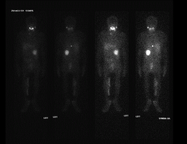
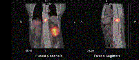
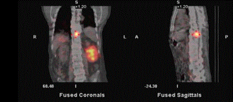
A 69 yr Vietnam war veteran with paraplegia and a history of PTC, s/p total thyroidectomy 5/13 (T3, N1) multifocal with lymphovascular and capsular invasion. 8/8 lymph nodes were positive and s/p I-131 remnant ablation 7/13 with 143 mCi. Suppressed Tg in 1/14 was 5.9 ng/mL. A dose of 254 mCi of I-131was given in 8/14 for metastatic PTC after L sided neck dissection and found 5/8 LN positive in 4/14 [1-3]. A post-RAI treatment scan revealed a focal uptake in the posterior component of T10//T11 vertebra which, in retrospect, was present in the prior post-ablation scan in 7/13 without any interval change. A chest CT in 9/14 revealed a sub centimeter metallic density in the same site, likely shrapnel deposited 45 years ago. Thus, focal radioiodine uptake likely relates to inflammatory/benign etiology. Suppressed Tg was 3.9 ng/mL in 8/15, decreased from 5.3 ng/mL in 2/15.
Figure 1
Figure 1
Post-treatment I-131 Total Body Scan on 8/19/2014: illustrates focal activity in the lower spine, at the
T10/T11 level which showed no interval change from a prior post-ablation scan on 7/24/2013.
Figure 2
Figure 3
Figure 4
Figure 4
CT of the Chest dated 9/29/2014 demonstrated a metallic density,
likely shrapnel posterior to the T10-T11 disc space and focal radioiodine
uptake likely relates to inflammatory/benign etiology.
Discussion
Focal radioiodine uptake is a sensitive marker for detection of recurrence of differentiated thyroid cancer, s/p total thyroidectomy and RAI ablation. However, radioiodine uptake is not specific for thyroid tissue. It can also be seen in healthy tissue, including thymus, breast, liver, and gastrointestinal tract, or in benign diseases, such as cysts and inflammation, or in a variety of benign and malignant non-thyroidal tumors, which could be mistaken for thyroid cancer. In order to accurately interpret radioiodine scintigraphy results, one must be familiar with the normal physiologic distribution of the tracer and frequently encountered physiologic and pathologic variants of radioiodine uptake. This case study provides another example of potential false-positive uptake of radioiodine in the whole body scan and illustrate how such unexpected findings can be appropriately evaluated [4]. Chronic trauma may recruit leukocytes that known to induce iodide organification by means of a myeloperoxidase. Therefore, retention of radioiodine in leukocytes of posttraumatic tissues may also explain various reports of false-positive uptake in sites of inflammation [5,6]. Secretion of mucin containing iodide salts has also been suggested as another possible mechanism of iodine accumulation associated with chronic inflammatory conditions.
Conclusion
Wounds caused by shrapnel fragments could be a common problem for veterans returning from overseas. Recognizing that false positive results could occur in RAI total body scan is clinically important.
References
- Wu SY, Brown T, Milne N. I-131 total body scan - extrathyroidal uptake of radioiodine, SeminNuc Med. 1986; 16(2): 82-84.
- Wu SY, Kollin J, Coodley E, Lockyer T, Lyons KP, Moran E, et al. Yu, I-131 total body scan: localization of disseminated gastric adenocarcinoma, J Nuc Med. 1984; 25(11): 1024-1029.
- Shen DH, Kloos RT, Mazzaferri EL, Jhian SM. Sodium iodide symporter in health and disease, Thyroid. 2001; 11(5): 415- 425.
- Oh JR, Ahn, BC. False-positive uptake on radioiodine whole-body scintigraphy: physiologic and pathologic variants unrelated to t hyroid cancer. Am J Nucl Med Mol Imaging. 2012; 2: 362-385.
- Klebanoff SJ, Hamon CB, Role of myeloperoxidase-mediated antimicrobial systems in intact leukocytes. J Reticuloendothel Soc. 1972; 12(2): 170-196.
- Regalbuto C, Buscema M, Arena S, Vigneri R, Squatrito S, Pezzino V. False-positive findings on (131)I whole-body scans because of posttraumatic superficial scabs. J Nucl Med. 2002; 43(2): 207-209.


