Review Article
Encapsulation of Doxorubicin in PLGA Nanoparticles Enhances Cancer Therapy
Lakshmi Priya Krishnamoorthy, Rajesh Kannan Moorthy, Devan Umapathy, Mahesh Kumar Kannan, Nithya Ganesan and Antony Joseph Velanganni Arockiam*
Department of Biochemistry, School of Life Sciences, Bharathidasan University, India
*Corresponding author: Antony Joseph Velanganni Arockiam, Department of Biochemistry, Bharathidasan University, India
Published: 14 Jun, 2017
Cite this article as: Krishnamoorthy LP, Moorthy RK,
Umapathy D, Kannan MK, Ganesan
N, Arockiam AJV. Encapsulation of
Doxorubicin in PLGA Nanoparticles
Enhances Cancer Therapy. Clin Oncol.
2017; 2: 1325.
Abstract
The anticancer effect of doxorubicin loaded PLGA nanoparticles on p-Dimethylaminoazobenzene
(p-DAB) induced liver cancer in male albino rats has been studied. Nanoparticles of poly (D,
L-Lactic-co-glycolic acid) (PLGA) were developed by nanoprecipitation method as delivery system
for doxorubicin. Doxorubicin was chemically conjugated to a terminal end group of PLGA. Group
I animals were fed with feed and water ad libitum and served as control. Group II-IV animals were
received p-DAB intraperitoneal injection at 20 mg/kg body weight once in a week for two months.
Then group III animals received intravenous injection of doxorubicin (240 μg/kg body weight) in
a tail vein daily. Group IV animals were received intravenous injection of PLGA nanoencapsulated
doxorubicin (420 μg of equivalent doxorubicin/kg body weight) in tail vein daily and stopped to the
conclusion of the experiment. Doxorubicin loaded PLGA nanoparticles were characterized by X-Ray
Diffractometer (XRD), Transmission Electron Microscopy (TEM) and Fourier Transmission Infra-
Red microscopy (FTIR). The elevated levels of protein, SGOT, SGPT in the p-DAB administered
group were significantly reduced by doxorubicin loaded PLGA nanoparticles than free doxorubicin.
In vivo anticancer activity showed that administration of doxorubicin loaded PLGA nanoparticles
had comparable activity to that of free doxorubicin.
Keywords: P-dimethylaminoazobenzene; Doxorubicin; Nanoparticles; liver cancer; Poly (D);
L-lactic-co-glycolic acid
Abbreviations
PLGA: Poly (D, L-Lactic-co-Glycolic Acid); P-DAB: P-Dimethylaminoazobenzene; XRD: X-ray
Diffractometer; TEM: Transmission Electron Microscopy; FTIR: Fourier Transmission Infra-Red
microscopy; PVA: Poly Vinyl Alcohol; SOD: Super Oxide Dismutase; SGPT: Serum Glutamate
Pyruvate Transaminase; SGOT: Serum Glutamate Oxaloacetate Transaminase; FDA: Food and
Drug Administration/p>
Introduction
Hepatocellular Carcinoma (HCC) is the major health problem of the world. Its incidence is increasing in both developing and developed countries. HCC represents the third cause of cancerrelated deaths [1]. P-Dimethylaminoazobenzene (p-DAB) is reported to induce damage in liver and caused tumors in several species of experimental animals by different route of administration [2]. Current cancer treatments can only prolong the life of the patients, but do not cure, because of the high toxicity and poor specificity of currently used drugs. Chemotherapeutic drugs are systemically active and cannot target cancer cells. Increasing the chemotherapeutic dosages is not possible. High dosage of chemotherapeutic drugs cause severe side effects like destruction of bone marrow cells which impairs the erythrocytes production, cardiotoxicity, nephrotoxicity, hepatotoxicity and hematotoxicity. To overcome these problems nanporticles are used in the cancer treatment. A nanoparticle-mediated drug delivery system can eliminate drug or drug carrier side effects significantly [3,4]. The major advantage of nanotechnology is targeted drug delivery to the site of the disease. This can be achieved either by passive targeting of drugs to the site of action or by active targeting of the drug [5]. Passive targeting exploits the anatomical differences between normal and diseased tissues to deliver the drugs to the required site, because the physiology of diseased tissues may be altered in a variety of physiological conditions through the enhanced permeability and retention (EPR) effect. The thermoplastic aliphatic polyesters like poly (lactide-co-glycolide) (PLGA), the copolymer of lactide and glycolide have generated tremendous interest due to their favorable properties such as good biocompatibility, biodegradability and mechanical strength [6]. They are easy to formulate into different devices for carrying a variety drug classes such as vaccines, peptides, proteins and micro molecules. Also, they have been approved by the Food and Drug Administration (FDA) for drug delivery [7]. Doxorubicin (Adriamycin) is a potent anti-neoplastic agent isolated from Streptomyces peuceticus. Doxorubicin was chemically conjugated to a terminal end group of PLGA by an ester linkage and the doxorubicin–PLGA conjugate was formulated into nanoparticles. A carboxylic acid end group of PLGA was conjugated to a primary hydroxyl group of doxorubicin. The primary amine group of doxorubicin was protected during the conjugation process and then deprotected. The nanoparticles containing the conjugate exhibited sustained doxorubicin release profiles over a month period [8]. Hence, the present work has been carried out to study the effect of doxorubicin-loaded PLGA on p-DAB induced liver cancer in rats.
Figure 1
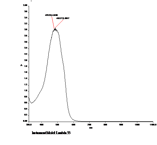
Figure 1
UV-Visible spectral peak of total drug.
Figure 2
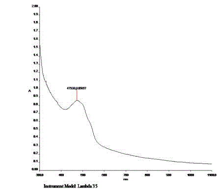
Figure 2
UV-Vis spectral peak of Free Drug.
Figure 3
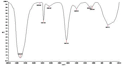
Figure 3
FTIR analysis of Free Drug.
Figure 4
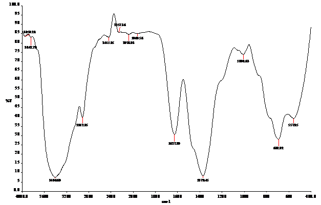
Figure 4
FTIR analysis of PLGA Polymer.
Figure 5
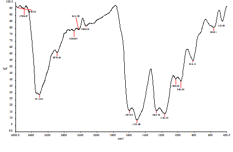
Figure 5
FTIR analysis of Doxorubicin loaded PLGA.
Figure 6
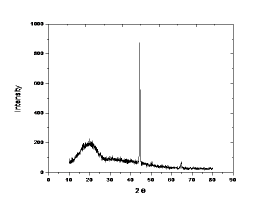
Figure 6
XRD analysis of Doxorubicin loaded PLGA.
Materials and Methods
Materials
Wistar male albino rats weighing 150 gm - 200 gm, reared
and maintained under the supervision of IAEC in the animal
house of the Bharathidasan University, were used for the study.
P-Dimethylaminoazobenzene (DAB) was purchased from LOBA
Chemie Pvt. Ltd, Mumbai-400 005. PLGA polymer was purchased
from Sigma Aldrich Co., 3050 Spruce Street, St.Louis, MO 63103
USA 314-771-5765. Polyvinyl alcohol (PVA) was purchased from
Hi Media Laboratories Pvt. Ltd, Mumbai-400 086. Doxorubicin
hydrochloride was purchased from Fresenius Kabi Oncology Ltd,
HP-173 205, India.
Experimental Design
All animals were acclimatized for 15 days and fed ad libitum
with rat feed and water. After acclimatization, animals were divided
into four groups consisting of 5 rats each. Group I animals were
served as normal control and received food and water for 90 days.
Group II animals were served as disease control and received DAB
(20 mg/Kg body weight) once in a week, intraperitoneally for 2
months [9,10]. Group III animals received DAB (20 mg/kg body
weight) intraperitoneally, once in a week for 2 months and after
received intravenous injection of doxorubicin hydrochloride (240 μg/
kg body weight) in a tail vein daily and stopped to the conclusion
of the experiment [11]. Group IV animals were received DAB (20
mg/kg body weight) intraperitoneally, once in a week for 2 months
and after received intravenous injection of PLGA nanoencapsulated
doxorubicin hydrochloride (420 μg of equivalent doxorubicin/kg
body weight) in tail vein daily and stopped to the conclusion of the
experiment.
Preparation of nanoparticles and encapsulation
The modified method of was used for nanoencapsulation [12].
The nanoprecipitation method was used for the formation of drugencapsulated
PLGA nanoparticles. PLGA polymer (40 mg) was
dissolved in acetone. Then the polymer solution containing same
amount of drug was added drop-wise into PVA aqueous solution
and stirred magnetically at room temperature until complete
evaporation of the organic solvent. Then, the nanoparticle suspension
was centrifuged by cooling centrifuge at 12,000 rpm for 1 hour. The
separated nanoparticles were redispersed and centrifuged three
times in distilled water to remove free drug completely. Finally,
nanoparticles were dried via vaccum drier at room temperature for 5
minutes, and then were characterized.
Table 1

Table 1
FTIR analysis of Functional Groups of Doxorubicin.
Table 2
Table 2
FTIR analysis of Functional Groups of PLGA Polymer.
Table 3
Table 3
FTIR analysis of Functional Groups of Doxorubicin loaded PLGA.
Results and Discussion
Evaluation of Doxorubicin Encapsulation
Nanoparticles were centrifuged at 12,000 g at 30 min in cooling
centrifuge, and then supernatants of Doxorubicin solutions were
measured by UV-spectrophotometer at 473 nm. Figure 1 and 2
showed the OD values of total drug (485.57) and free drug (473.10).
Calculations were performed by using the calibration curve, and
encapsulation efficiency was calculated as described [13].
Doxorubicin encapsulation efficiency (%) = Total Doxorubucin −
Free Doxorubicin X 100/Total Doxorubucin
= 20 mg – 5.0 mg X 100/20 mg
= 75
In the present study, Doxorubicin encapsulation efficiency was
as 75 for 20 mg of PLGA polymer. However, that the encapsulation
efficiency of chitosan nanoparticles (5.0 mg/mL 5-FU) was 29.98 ±
0.8 [14].
FTIR Analysis
Infrared spectroscopy was used to study the interactions between
the drug and the polymers. In order to investigate possible molecular
interaction between drug and the polymer, FTIR spectroscopy
(Perkin-Elmer, Spectrum RXI) of the Doxorubicin, PLGA polymer
and Doxorubicin-loaded PLGA nanoparticles (NPs) was carried out.
The FTIR spectra were collected in the range of 4000 cm-1 – 400 cm-1
at room temperature. FTIR analysis of free drug (Figure 3) showed
distinct peaks at wave numbers 667.73 cm-1, 1031.20 cm-1, 1098.86 cm-
1, 1400.12 cm-1, 1637.12 cm-1, 2085.67 cm-1, 2357.89 cm-1, 3445.65 cm-
1. The peaks corresponding to Alkyne C-H, Aromatic C-H, Aromatic
C-H, Carboxylic acids, Alkenyl C=C stretch, NH3 structure, Charged
amines C=NH+, H-bonded OH stretch (Table 1).
FTIR analysis of PLGA polymer (Figure 4) showed distinct peaks
at wave numbers 692.02 cm-1, 1008.63 cm-1, 1370.45 cm-1, 1627.90 cm-
1, 1960.56 cm-1, 2257.36 cm-1, 2441.85 cm-1, 3406.60 cm-1. The peaks
corresponding to thioether CH2-S, cyclohexane ring, methyl C-H
bend, alkenyl C=C stretch, aromatic combination bands, saturated
nitriles C≡N, charged amines C=NH+, OH stretch (Table 2).
FTIR analysis of Doxorubicin loaded PLGA (Figure 5) showed
distinct peaks 963.28 cm-1, 1026.05 cm-1, 1164.52 cm-1, 1263.78 cm-1,
1501.89 cm-1, 1597.23 cm-1, 2266.65 cm-1, 2451.92 cm-1, 2566.65 cm-1,
2976.48 cm-1, 3414.25 cm-1. The peaks corresponding to trans C-H
stretch, cyclohexane ring vibrations, aromatic C-H, esters of aromatic
acid, aromatic ring stretch, aromatic ring stretch, diazonium salts
(R-C=N=N)+, charged amines C=NH+, thiols S-H stretch, alkanes –
CH3, H bonded OH stretch. Doxorubicin was chemically conjugated
to a terminal end group of poly (D, L-lactic-co-glycolic acid) [PLGA]
by an ester linkage. 1263.78 cm-1 showed that ester linkage (Table 3
and 4) [15]. Reported that the spectra of the PLGA NPs and Alpha
1-antitrypsin-loaded PLGA NPs showed two distinct peaks at wave
numbers 1300 cm-1 - 1450 cm-1 and 1720 cm-1 - 1800 cm-1, which were
assigned to δCH and V(C=O).
Figure 7
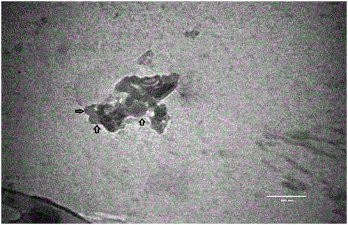
Figure 7
Transmission Electron Microscopy of Doxorubicin loaded PLGA.
Figure 8
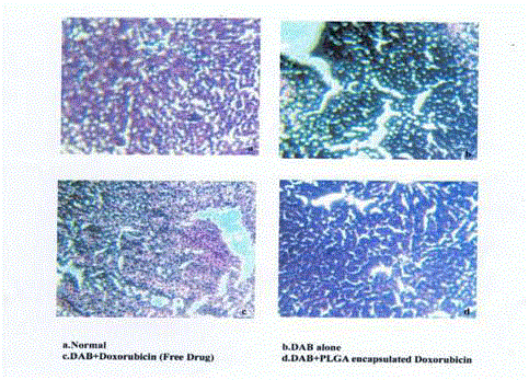
Figure 8
Histopathological Appearance of Rat Liver.
Figure 9
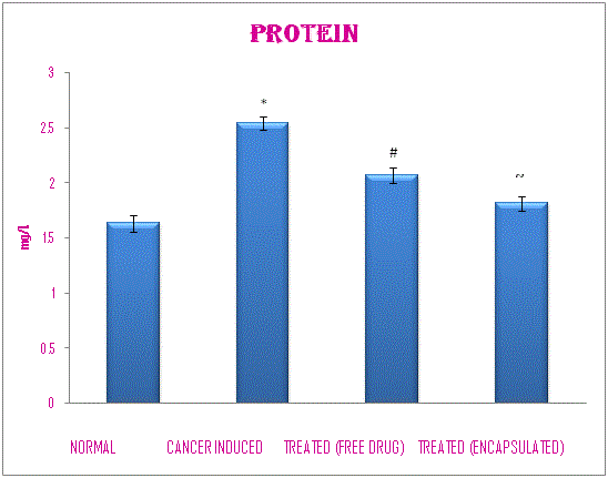
Figure 9
Effect of Doxorubicin on Protein Levels of Rats Administered With
p-DAB.
XRD Analysis
Showed the diffraction lines Figure 6 and 7, which were observed at 2θ angle 16.780°, 20.440° and 21.100° and 23.060°. By using 2θ angle, the size of the nanoparticle was calculated. Debye-Scherrer’s formula was used to calculate the size of the nanoparticle [16]. Reported that the XRD pattern of pure silver ions was known to display peaks at 2θ= 7.9°, 11.4°, 17.8°, 30°, 38°, and 44°.
Figure 10
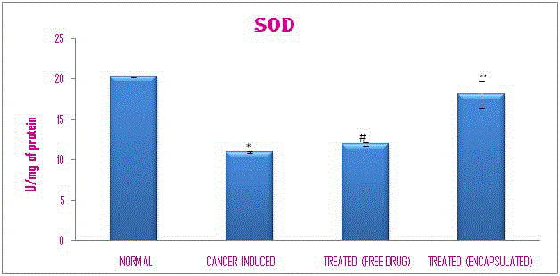
Figure 10
Effect of Doxorubicin on Super Oxide Dismutase Levels of Rats
Administered with p-DAB.
Figure 11
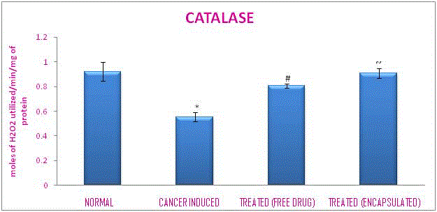
Figure 11
Effect of Doxorubicin on Catalase Levels of Rats Administered
With p-DAB.
Figure 12
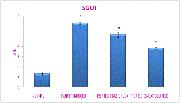
Figure 12
Effect of Doxorubicin on SGOT Levels of Rats Administered With
p-DAB.
Figure 13
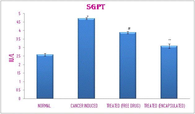
Figure 13
Effect of Doxorubicin on SGPT Levels of Rats Administered Wit
p-DAB.
TEM Analysis
Microphotographs of the Doxorubicin loaded PLGA were obtained by transmission electron microscopy (Zeiss). Figure 7 showed the Transmission Electron Microscope (TEM) image of the Doxorubicin loaded PLGA nanoparticles. It revealed that the NPs have spherical shape. The resulting NPs were predominantly spherical and of uniform size and shape [17]. Reported that the imaging of the GC–BTNPs by TEM revealed well-dispersed structures [18]. Reported that the TEM of freeze- fractured samples showed that the particles had a central cavity surrounded by a polymer wall.
Table 4
Table 4
Measurement of the Doxorubicin loaded PLGA nanoparticles by using
Debye-Scherrer’s equation.
Table 5
Table 5
Effect of Doxorubicin on Protein Levels of Rats Administered with p-DAB.
Table 6
Table 6
Effect of Doxorubicin on Super Oxide Dismutase levels of rats
administered with p-DAB.
Table 7
Table 7
Effect of Doxorubicin on Catalase Levels of Rats Administered With
p-DAB.
Table 8
Table 8
Effect of Doxorubicin on SGOT Levels of Rats Administered with p-DAB.
Table 9
Table 9
Effect of Doxorubicin on SGPT Levels of Rats Administered with p-DAB.
Histopathological Appearance of Rat Liver
Normal rats show more compact and well-distributed junctional complexes (Figure 8a). In DAB-administered rats, hepatic cells exhibit damaged cell structure (Figure 8b). However, animals treated with doxorubicin (Figure 8c) show high cell density with compact junctional complexes than DAB-administered animals. The animals treated with doxorubicin loaded PLGA show higher cell density with compact junctional complexes (Figure 8d) than doxorubicin treated animals. Velanganni and Balasundaram reported that hepatic cells exhibit loss of contact inhibition (polarity) and damaged central vein of liver lobules in DAB-administered rats (Figure 9).
Biochemical Analysis
The Figure 9 showed the effect of doxorubicin on protein levels
of rats administered with p-DAB. Protein level was estimated by
Lowry’s method [19]. Protein level, which increased after DAB
administration, was found to be lowered significantly (P <0.001) by
Doxorubucin loaded PLGA when compared to free Doxorubicin
(Table 5). Figure 10 and 11 showed the effect of doxorubicin on SOD
and catalase levels of rats administered with p-DAB respectively. SOD
was estimated [20,21]. Catalase level was estimated by Sinha’s method
[22]. Antioxidants SOD and catalase, which were decreased after DAB
administration and found to be increased after the administration of
Doxorubicin and Doxorubicin loaded PLGA. Doxorubicin loaded
PLGA increased the antioxidants levels significantly (P <0.001; P
<0.005) when compared to free doxorubicin (Table 6 and 7).
Figure 12 and 13 showed the effect of doxorubicin on SGOT and
SGPT levels of rats administered with p-DAB respectively. SGOT and
SGPT levels were estimated by Reitman’s method. SGOT and SGPT
levels, which increased after DAB administration, were found to be
lowered significantly (P <0.005) by Doxorubicin loaded PLGA when
compared to free Doxorubicin. Reported that the levels of GSH, ALP,
GST and bilirubin were increased after DAB administration and these
were found to be lowered by vitamins A, C and E (Table 8 and 9).
Conclusion
The in vivo anti-tumor activity of the PLGA loaded doxorubicin nanoparticle was higher than the free doxorubicin. Hence, Doxorubicin loaded PLGA nanoparticle can be used to improve the therapeutic efficacy of Doxorubicin in the treatment of cancer.
Acknowledgement
Ms. K. Lakshmi Priya is thankful to DST for the award of INSPIRE Fellowship and DST-FIST Sponsored Dept. of Biochemistry, Bharathidasan University, Tiruchirappalli for providing the platform to do the research work.
References
- Arjmand Sareh, Elham Bidram, Abbas Sahebghadam Lotfi, Hamid Mahdavi. Evaluation of Poly (D, L-lactide-co-glycolide) for Nanoencapsulation of Alpha 1-antitrypsin and In Vitro Release Study, International Journal of Bioscience, Biochemistry and Bioinformatics. 2011.
- Aydin Seda Tigl R, Mehlika Pulat. 5-Fluorouracil Encapsulated Chitosan Nanoparticles for pH-Stimulated Drug Delivery: Evaluation of Controlled Release Kinetics, J Nanomaterials. 2012.
- Cheung TK, Lai CL, Wong B CY, Fung J, Yuen MF. Clinical features, biochemical parameters, and virological profiles of patients with hepatocellular carcinoma in Hong Kong. Aliment Pharmacol Ther. 2006; 24(4): 573-583.
- Ciofani Gianni, Serena Danti, Delfo D’Alessandro, Stefania Moscato, Mario Petrini, Arianna Menciassi. Barium Titanate Nanoparticles: Highly Cytocompatible Dispersions in Glycol-chitosan and Doxorubicin Complexes for Cancer Therapy, Nanoscale Research Letter. 2010; 5(1): 1093-1101.
- Derakhshandeh K, Erfan M, Dadashzadeh S. Encapsulation of 9-nitrocamptothecin, a novel anticancer drug, in biodegradable nanoparticles: factorial design, characterization and release kinetics. Eur J Pharm Biopharm. 2007; 66(1): 34-41.
- Derakhshandeh Katayoun, Marzieh Soheili, Simin Dadashzadeh, Reza Saghiri. Preparation and in vitro characterization of 9-nitrocamptothecin-loaded long circulating nanoparticles for delivery in cancer patients. Int J Nanomedicine. 2010; 5: 463-471.
- Dubey Manish, Seema Bhadauriaa, Kushwahb BS. Green synthesis of nano silver particles from extract of Eucalyptus hybrida (Safeda) leaf, Digest Journal of Nanomaterials and Biostructures. 2009; 4(2): 537-543.
- Yoo HS, Lee KH, Oh JE, Park TG. In vitro and in vivo anti-tumor activities of nanoparticles based on doxorubicin-PLGA conjugates. J Control Release. 2000; 68(3): 419-431.
- Hyuk Sang Yoo, Keun Hyeung Lee, Jong Eun Oh, Tae Gwan Park. Biodegradable Nanoparticles Doxorubicin-PLGA conjugate for Sustained Release. Pharm Res. 1999; 16(7): 1114-1118.
- IARC. Para-Dimethylaminoazobenzene. In Some Aromatic Azo Compounds. IARC Monographs on the Evaluation of Carcinogenic Risk of Chemicals to Humans, vol. 8. Lyon, France: International Agency for Research on Cancer. 1975; 125-146.
- Jin S, Ye K. Nanoparticle-mediated drug delivery and gene therapy. Biotechnol Prog. 2007; 23(1): 32-41.
- Kumar CSSR, Hormes J, Leuschner (Eds). Nanofabrication Towards Biomedical Applications, Techniques, Tools, Applications, and Impact, Weinheim, WILEY-VCH Verlag GmbH&Co.KGaA. 2005.
- Lowry OH, Rosebrough NJ, Farr AL, Randall RJ. Protein measurement woth the Folin-Phenol reagents, J Biol Chem. 1951; 193(1): 265-275.
- Markland S, Markland G. Involvement of the superoxide anion radical in the auto oxidation of pyrogallol and a convenient assay for super oxide dismutase, Eur J Biochem. 1974; 47(3): 469-474.
- Jain RA. The manufacturing techniques of various drug loaded biodegradable poly(lactide-co-glycolide) (PLGA) devices. Biomaterials. 2000; 21(23): 2475-2490.
- Reitman S, Frankel S. In vitro determination of transaminases in serum, Am J Clin Pathol. 1957; 28(1): 56-63.
- Sinha AK. Colorimetric assay of catalase. Anal Biochem. 1972; 47(2): 389-394.
- Suphiya Parveen, MS, Ranjita Misra, MS, Sanjeeb K. Sahoo Nanoparticles: a boon to drug delivery, therapeutics, diagnostics and imaging, Nanomedicine. Nanomedicine. 2012; 8(2): 147-166.
- Velanganni AAJ, Balasundaram C. Protective effect of vitamin A, ascorbic acid and Æ-tocopherol on 2, 4-dimethylaminoazobenzene-induced hepatoma in rats. Current Science. 2013; 85(2).
- Watnasirichaikul Suchat, Nigel M, Davies, Thomas Rades, Ian G. Tucker Preparation of Biodegradable Insulin Nanocapsules from Biocompatible Microemulsions. Pharma Res. 2000; 17(6).
- WHO, Monographs on the evolution of carcinogenic risk of chemicals to man. 1975; 8.