Editorial
Diagnostic Pitfalls in Axillary Sentinel Lymph Node Evaluation
Emmanuel Agosto-Arroyo and Marilin Rosa*
Department of Anatomic Pathology, Moffitt Cancer Center, USA
*Corresponding author: Marilin Rosa, Department of Anatomic Pathology, Comprehensive Breast Program, Moffitt Cancer Center, 12902 Magnolia Drive, Tampa, FL 33612, USA
Published: 21 Dec, 2016
Cite this article as: Agosto-Arroyo E, Rosa M. Diagnostic
Pitfalls in Axillary Sentinel Lymph Node
Evaluation. Clin Oncol. 2016; 1: 1165.
Introduction
Surgical excision of axillary lymph node(s) is standard practice in the management of invasive breast cancer. Whether sentinel or axillary lymph node excisions are performed, typically the histopathological distinction of metastatic carcinoma is straightforward. However, there are rare, though, important pathological entities that may pose a diagnostic challenge in the evaluation of axillary sentinel lymph nodes, including heterotopic epithelial inclusion and nevus cell aggregates.
Heterotopic Epithelial Inclusions
In the axilla, heterotopic inclusions can be found within the lymph node capsule and/or
parenchyma [1-4]. The morphologic features of these inclusions are varied. Heterotopic mammarytype
glands can be present in the axillary lymph node and resemble benign mammary epithelium, as
well as, undergo fibrocystic changes, epithelial hyperplasia, apocrine metaplasia, cysts and sclerosing
adenosis [5]. Mammary-type glands are the most common heterotopic inclusion in the axillary
lymph nodes [6]. Other types of inclusions include: squamous epithelium and mixed squamous
and glandular components. Glandular elements with müllerian-like features can also be seen in
a minority of axillary lymph nodes heterotopic inclusions, typically identified by the presence of
microvilli [1].
The presence of heterotopic epithelial inclusions in the axillary lymph node is rare and the origin
is unknown, but displaced epithelium during prior procedure [5] and embryologic maldevelopment
[6] have been hypothesized as possible histogenesis.
The differential diagnosis of heterotopic epithelial inclusions includes metastatic carcinoma to
the lymph node. The presence of glandular elements within the nodal capsule and intraparencymal,
rather than the lymph node sinusoids, is suggestive of heterotopic epithelial inclusions. Most of
the heterotopic glandular elements of mammary-type show evidence of myoepithelial cells by
Hematoxylin and Eosin (H&E) and with myoepithelial cell markers on immunohistochemistry.
This fact does not hold true for inclusions with squamous and müllerian-like features, both of which
lack evidence of myoepithelial cell markers. Inclusions of müllerian-like origin are immunoreactive
for WT-1 and PAX-8 [7].
Nevus Cell Aggregates
Melanocytic nevus presenting as intracapsular and intratrabecular cell aggregates is a wellknown
diagnostic entity in various anatomic locations. The cells forming the nevus aggregates
resemble intradermal nevi, ranging from oval, or spindle cell, to epithelioid cells. Some aggregates
may lack or contain minimal pigment, others may show substantial pigment content, resembling
blue nevi. Their location is commonly intracapsular, however, intratrabecular and intraparenchymal
nevus cell aggregates can arise within the lymph node.
Nevus cell aggregates occur more frequently in the axilla than in other anatomic locations and
have been found in 0.54% of examined axillary lymph nodes [8]. The histogenesis of nevus cell
aggregates is unknown. A possible etiology is that the cells are derived from perithelial cells around
the nodal vasculature [9]. Another possible etiology is the development of the nevus cell aggregates
from embryologic remnants.
Metastatic carcinoma can easily be ruled out from the differential diagnosis of nevus cell
aggregates by lack of immunohistochemical staining with pancytokeratin. Metastatic melanoma
and nevus cell aggregates are both positive to S100, MART-1 and Melan-A. HMB45 is positive in
metastatic melanoma and negative in nevus cell aggregates [10]. Another useful marker is Ki-67, which has been reported to be less than 1% in nevus cell aggregates [10] and highly positive in metastatic carcinoma.
Conclusion
The presence of metastatic carcinoma within the lymph node is a marker of loco-regional disease and an independent prognostic factor. Accomplishing the correct pathological diagnosis is of utmost importance management of patients with invasive breast cancer. Awareness of rare, but benign, lesions within the axillary lymph node helps the pathologist to avoid misinterpretation with metastatic neoplasms.
Figure 1
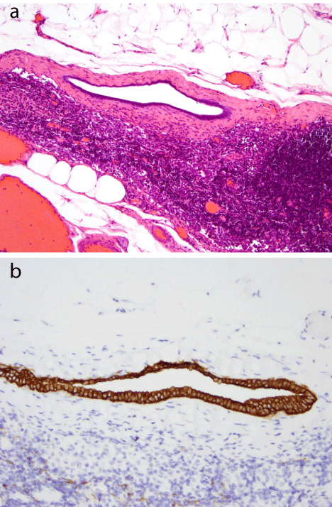
Figure 1
Intracapsular heterotopic epithelial inclusion with a lining of single
cells with focal pseudostratification (a, H&E 100X), showing positivity to
cytokeratin AE1/AE3 immunohistochemical stain (b, IHC 200X).
Figure 2
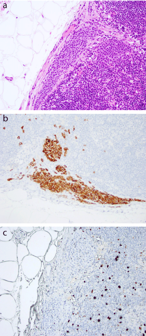
Figure 2
Subcapsular nevus cell aggregate (a, H&E 200X), with
infiltration of the cells into the lymph node parenchyma as seen by the
immunohistochemical stain with Melan-A (b, IHC 200X). The nevus cell
aggregates are negative to Ki-67 proliferation index (c, IHC 200X).
References
- Holdsworth PJ, Hopkinson JM, Leveson SH. Benign axillary epithelial lymph node inclusions—a histological pitfall. Histopathology. 1988; 13: 226–228.
- Layfield LJ, Mooney E. Heterotopic epithelium in an intramammary lymph node. Breast J. 2000; 6: 63–67.
- Fellegara G, Carcangiu ML, Rosai J. Benign epithelial inclusions in ax-illary lymph nodes: report of 18 cases and review of the literature. Am J Surg Pathol. 2011; 35: 1123–1133.
- Zynger DL, McCallum JC, Everton MJ, Yeldandi AV, Susnik B. Paracortical axillary sentinel lymph node ectopic breast tissue. Pathol Res Pract. 2009; 205: 427–432.
- Hoda SA, Brogi E, Koerner FC, Rosen PP. Rosen’s Breast Pathology (4th Ed.). Philadelphia, PA. LWW. 2014.
- Resetkova E, Hoda SA, Clarke JL, Rosen PP. Benign heterotopic epithelial inclusions in axillary lymph nodes: histological and immunohisto-chemical patterns. Arch Pathol Lab Med. 2003; 127: e25–e27.
- Corben AD, Nehhozina T, Garg K, Vallejo CE, Brogi E. Endosalpingiosis in axillary lymph nodes: a possible pitfall in the staging of patients with breast carcinoma. Am J Surg Pathol. 2010; 34: 1211–1216.
- Bautista NC, Cohen S, Anders KH. Benign melanocytic nevus cells in axillary lymph nodes. A prospective incidence and immunohistochem-ical study with literature review. An J Clin Pathol. 1994; 102: 102–108.
- Micheau C, Contesso G. Formations d’aspect angioglomique dans les ganglions lymphatiques axillaires. Arch Anat Pathol. 1971; 19: 167–175.
- Biddle DA, Evans HL, Kemp BL, El-Naggar AK, Harvell JD, White WL, et al. Intraparenchymal nevus cell aggreagtes in lymph nodes-A possible diagnostic pitfall with malignant melanoma and carcinoma. Amm J Surg Pathol. 2003; 27: 673-681.