Clinical Image
Ga-68 PSMA Accumulation in Hepatocellular Carcinoma
Soydal C1*, Alkan A2, Ozkan E1, Demirkazık A2 and Kucuk NO1
1Departments of Nuclear Medicine, Ankara University, Turkey
2Departments of Medical Oncology, Ankara University, Turkey
*Corresponding author: Cigdem Soydal, Ankara University Medical School, Department of Nuclear Medicine, 06590, Cebeci, Ankara, Turkey
Published: 12 Sep, 2016
Cite this article as: Soydal C, Alkan A, Ozkan E,
Demirkazık A, Kucuk NO. Ga-68
PSMA Accumulation in Hepatocellular
Carcinoma. Clin Oncol. 2016; 1: 1091.
Abstract
We present intense uptake in Ga-68 prostate specific membrane antigen (PSMA) positron emission
tomography (PET)/computed tomography (CT) in primary hepatocelullar carcinoma (HCC) as
well as its metastases in 72 years old male patient, who have prostate, bladder and HCC carcinoma
diagnoses.
Keywords: Ga-68 prostate specific membrane antigen; Positron emission tomography/
Computed tomography; Hepatocellular carcinoma
Clinical Image
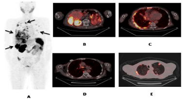
A 72 years old male patient underwent Ga-68 PSMA PET/CT for restaging of prostate carcinoma. He had diagnosed as prostate adenocarcinoma in 2010 and been followed up with bone metastases under androgen deprivation and zoledronic acid therapies. Additionally segmental liver resection and radiofrequency ablation therapy had been administered in January 2015 for HCC. Finally chemoradiation therapy following partial cystectomy had been applied for high grade invasive bladder carcinoma. After chemotherapy and sorafenib treatment, patient underwent Ga- 68 PSMA PET/CT for elevation of serum prostate specific antigen levels. In maximum intensity projection images (A) and transaxial fused images, pathological uptake was detected in liver lesions (B), pleural surface in right hemithorax (C), mediastinal lymph nodes (D) and nodules in both lung (E). After PET/CT patient underwent fine needle aspiration biopsy from liver mass and pleura and hepatocellular carcinoma metastases were confirmed by histopathological examination. Ga- 68 PSMA PET/CT has been performed for imaging of recurrent prostate carcinoma [1]. Despite specific binding of PSMA molecule for prostate carcinoma cell surface, false positive uptake in renal cell carcinoma, breast cancer and thyroid cancer has been reported as case reports [2-4]. Ga-68 PSMA uptake in HCC has been reported in only one case by Sasikumar et al. [5] recently. In this case we would like to share our experience of intense Ga-68 PSMA uptake in our metastatic HCC patient.
Figure
Figure
References
- Afshar-Oromieh A, Avtzi E, Giesel FL. The diagnostic value of PET/CT imaging with the (68)Ga-labelled PSMA ligand HBED-CC in the diagnosis of recurrent prostate cancer. Eur J Nucl Med Mol Imaging. 2015; 42: 197-209.
- Sager S, Vatankulu B, Uslu L. Incidental Detection of Follicular Thyroid Carcinoma in Ga-68 PSMA PET/CT Imaging. J Nucl Med Technol. 2016; 3: 10.
- Demirci E, Ocak M, Kabasakal L. (68)Ga-PSMA PET/CT imaging of metastatic clear cell renal cell carcinoma. Eur J Nucl Med Mol Imaging. 2014; 4: 1461-1462.
- Sathekge M, Modiselle M, Vorster M. 68Ga-PSMA imaging of metastatic breast cancer. Eur J Nucl Med Mol Imaging. 2015; 42: 1482-1483.
- Sasikumar A, Joy A, Nanabala R. (68)Ga-PSMA PET/CT imaging in primary hepatocellular carcinoma. Eur J Nucl Med Mol Imaging. 2016; 43: 795-796.