Case Report
Tuberculosis of the Breast
Orerah GI1* and Wasike RW2
1Department of Surgery, Aga Khan University Hospital, Kenya
2Department of Head of Breast Service, Aga Khan University Hospital, Pakistan
*Corresponding author: George Isaya Orerah, Department of Surgery, Aga Khan University Hospital, PO Box 62950 00200 Nairobi, Kenya
Published: 02 Sep, 2016
Cite this article as: Orerah GI, Wasike RW. Tuberculosis of
the Breast. Clin Oncol. 2016; 1: 1068.
Abstract
Tuberculosis of the breast is an extremely uncommon presentation of an otherwise common disease
especially in developing countries. Clinical presentation is usually of a solitary, ill-defined, unilateral
hard lump situated in the upper outer quadrant of the breast and may mimic breast carcinoma
or breast abscess. Appropriate diagnosis precludes surgery as medical management suffices. We
present two cases of this rare presentation that highlight the peculiarities of tuberculosis of the
breast as well as a review of the literature.
Keywords: Breast disease; Breast tuberculosis
Introduction
Tuberculosis is the most widespread and persistent human infection in the world. The infection
can involve any organ and mimic other illness; hence it is called the great mimicker.
Breast tuberculosis (TB) was first defined by Sir Astley Cooper in 1829 [1]. It is an extremely
uncommon disease responsible for 0.025-1.04% of all breast pathologies [2]. It is an uncommon
disease even in countries where the incidence of tuberculosis is high.
The disease is very rare in males. In a review by Gupta et al.[3] comprising 160 patients, only 6
were males. In Japan, only two cases were reported in males out of 52 cases of mammary tuberculosis
over a period of 15 years from 1986 to 2000 [4].
In this article we report a case of male breast tuberculosis in a 70 year old alongside another case
of a lady with tuberculous abscess.
Case Presentation
Case 1
A 70 year old man presented with a 6-month history of a lump in his left breast, associated with
mild pain, fever, night sweats, loss of appetite and weight loss. He had a history of lymphoma 5 years
prior (2009) for which he underwent chemotherapy and cervical lymph node dissection. There was
no family history of tuberculosis.
On examination, he was in obvious respiratory distress as evidenced by inability to complete full
sentences and tachypnea. He had reduced breath sounds and crepitations evident on his right mid
and lower lung zones.
On breast examination, there was an obvious left breast mass, estimated 4 by 4 cm that was
centrally located. The mass was firm with minimal tenderness. It was not mobile and was attached
to the underlying tissues but not the overlying skin. There were no skin or nipple changes. No nipple
discharge was evident.
Patient was sent for an ultra-sound guided aspiration and core needle biopsy of the mass. PCR
mycobacterium identification and sensitivity was positive. With this a diagnosis of tuberculosis of
the breast was made.
The patient was placed on a regimen of anti-tuberculosis quadritherapy with significant
improvement a month later. He is still on treatment and follow-up.
Case 2
A 58 year old postmenopausal lady known to have diabetes and hypertension. She presented
with complaints of right breast lump for six weeks. The lump had been slowly increasing in size
with associated discomfort. She had nipple discharge of whitish and sometimes bloody fluid one
week prior to her presentation. She had no other symptoms like fever, weight loss, loss of appetite
or malaise. On examination, she had a right lower inner quadrant
breast mass measuring 4 by 4 cm with moderate tenderness and
cystic consistency. There was nipple retraction and erythema around
the mass. She did not have lymphadenopathy. Her other systemic
examination were all normal. An impression of a right breast abscess
was made and she was taken for an incision and drainage procedure.
Samples were taken for microscopy, culture and sensitivity. The
cultures turned positive for granulomatous inflammation suspicious
of mycobacterium tuberculosis. She was promptly referred to the
tropical medicine specialists for initiation of anti TB therapy and
follow up. She also continued with dressing changes of the wound
which has healed well.
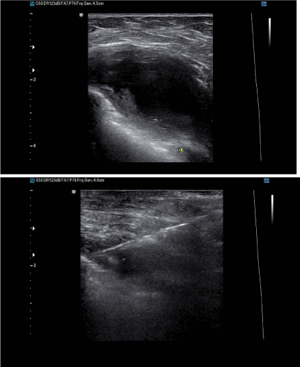
Breast ultra-sound done showed hypoechoeic left breast mass 56*45*36
mm.
Discussion
TB remains one of the leading causes of death from infectious
diseases worldwide. Breast TB is one of its rarest forms [5]. The
first case was recorded by Sir Astley Cooper in 1829 who called it
‘scrofulous’ swelling of the bosom [6]. It comprises 3% of all breast
diseases and is 5 times less common than carcinoma of the breast
[7]. It can be classified as primary when no demonstrable tuberculous
focus exists, or it can be secondary to a lesion elsewhere in the body
[8,9].
Breast TB almost exclusively affects women and occurs mostly in
multiparous lactating women between the ages of 20 and 40 years [10].
This parallels the highest incidence of pulmonary tuberculosis [11].
This could be because the female breast undergoes frequent changes
during the period of childbearing activity and is more susceptible to
trauma and infection [12].
The disease is very rare in males. In India, a review by Gupta et
al. [3] comprising 160 patients, only 6 were males comprising 3.75%.
In Japan, only two cases were reported in males out of 52 cases of
mammary tuberculosis over a period of 15 years from 1986 to 2000
[4]. In this case presentation, it occurred in a male of 70 years. Clinical
presentation is usually of a solitary, ill-defined, unilateral hard lump
situated in the central or upper outer quadrant [11,13-15], similar to
our case. TB can also present with nipple discharge, skin thickening,
or discharging sinuses in the breast or axilla, seen in our second case
presentation. The lesion may be indistinguishable from carcinoma
breast, being irregular, hard, and at times, fixed to either skin or
muscle or even chest wall [16]. Pulmonary involvement occurs only
rarely [13]. Constitutional symptoms of fever, anorexia, failing health,
weight loss and night sweats have been reported before [17] and was
in our patient as well.
It is generally thought that the breast gets involved in tuberculosis
by retrograde lymphatic extension from the mediastinal, axillary, and
cervical region [18]. This hypothesis is supported by the involvement
of axillary nodes, frequently ipsilateral nodes, in 50% to 75% of
tuberculous mastitis cases [16,19].
Breast TB can mimic breast carcinoma or breast abscess,
clinically and radiographically [5,15]. In our case, the lady presented
with a breast abscess. Radiological imaging is not diagnostic.
Mammographic imaging may show a dense tract connecting an illdefined
breast mass to an area of skin thickening and a skin bulge.
Ultrasound may demonstrate a complex, predominantly cystic mass.
Diagnosis is based on identification of typical histological features
or the tubercle bacilli under microscopy or culture. Shah et al. [20]
performed AFB smear, AFB culture, and the DAT (Amplicor assay)
on 1090 tissue and body fluid specimens. They found PCR test to be
very useful for detecting M. tuberculosis in non-respiratory samples,
which have lower frequency of positive AFB smear [21].
Medical therapy is the mainstay of therapy with anti-tuberculous
therapy. Surgical intervention was needed in up to 14% of the patients
in some series, either due to lack of response to chemotherapy or
large painful ulcerative lesions involving the entire breast [22,23].
Simple mastectomy is rarely needed now-a-days and is reserved for
patients with extensive disease comprising large painful ulcerated
mass involving the entire breast and draining axillary lymph nodes.
He had a needle aspirate microscopy which was negative for acid fast bacilli
(ZN staining).
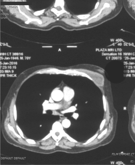
CT scan of the chest showed a left chest wall soft tissue mass measuring
31mm*56mm*54mm which was attached to the chest wall muscles. The
overlying breast tissue was of normal attenuation.
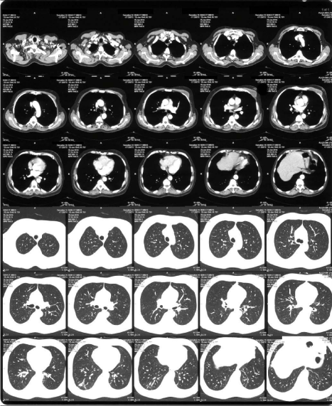
The lung parenchyma appeared normal.
Conclusion
Tuberculosis of the breast is rare and tuberculosis of the male breast is not a recognised entity. The clinical and radiological feature of mammary tuberculosis can be very confusing and easily mistaken for breast cancer. Symptoms suggestive of tuberculosis warrant a biopsy to exclude possible cancer. Incorporating a highly sensitive technique like polymerase chain reaction (PCR) may be helpful in establishing the usefulness of such technology and can aid in confirming the diagnosis early [24]. The disease is curable with antitubercular drugs, and surgery is rarely required.
References
- Cooper A. Illustration of the Diseases of the Breast. Part I. London: Longman, Rees, Orme, Brown and Green; 1829. 7.
- Bani-Hani KE, Yaghan RJ, Matalka II, Mazahreh TS. Tuberculosis mastitis: a disease not to be forgotten. Int J Tuberc Lung Dis. 2005; 9: 920-925.
- Gupta PP, Gupta KB, Yadav RK, Agarwal D. Tuberculous mastitis: A review of seven consecutive cases. Indian J Tub. 2003; 50: 47-50.
- Khanna R, Presanna G V, Gupta P, Kumar M, Khanna S, Khanna A K. Mammary tuberculosis: report on 52 cases. Postgrad Med J. 2002; 78: 422-424.
- Sen M, Gorpelioglu C, Bozer M. Isolated primary breast tuberculosis: report of three cases and review of the literature. Clinics (Sao Paulo). 2009; 64: 607-610.
- Cooper A. Illustrations of the Diseases of the Breast, Part I. London: Longman, Rees, Orme, Brown and Green; 1829. 73.
- Khanna R, Prasanna GV, Gupta P, Kumar M, Khanna S, Khanna AK. Mammary tuberculosis: report on 52 cases. Postgrad Med J. 2002; 78: 422-424.
- Shinde SR, Chandawarkar RY, Deshmukh SP. Tuberculosis of the breast masquerading as carcinoma: a study of 100 patients. Word J Surg. 1995; 19: 379-381.
- Kakker S, Kapila K, Singh MK, Verma K. Tuberculosis of the breast. A cytomorphologic study. Acta Cytol. 2000; 44: 292-296.
- Elsiddig KE, Khalil EA, Elhag IA, Elsafi ME, Suleiman GM, Elkhidir IM, et al. Granulomatous mammary disease: ten years' experience with fine needle aspiration cytology. Int J Tuberc Lung Dis. 2003; 7: 365-369.
- Jaideep C, Kumar M, Klianna AK. Male breast tuberculosis. Postgrad Med J. 1997; 73: 428-429.
- Raw N. Tuberculosis of the breast. Br Med J. 1924; 1: 657-658.
- Jalali U, Rasul S, Khan A, Baig N, Khan A, Akhter R. Tuberculous mastitis. J Coll Physicians Surg Pak. 2005; 15: 234-237.
- Banerjee A, Green B, Burke M. Tuberculous and granulomatous mastitis. Practitioner. 1989; 233: 754-756.
- Baharoon S. Tuberculosis of the breast. Ann Thorac Med. 2008; 3: 110.
- Shinde SR, Chandawarkar RY, Deshmukh SP. Tuberculosis of the breast masquerading as carcinoma: A study of 100 patients. World J Surg. 1995; 19: 379-381.
- Bouti K, Soualhi M, Marc K, Zahraoui R, Benamor J, Bourkadi JE, et al. Postmenopausal Breast Tuberculosis-Report of 4 cases. Breast Care (Basel). 2012; 7: 411-413.
- Sen M, Gorpelioglu C, Bozer M. Isolated primary breast tuberculosis: report of three cases and review of the literature. Clinics (Sao Paulo) 2009; 64: 607-610.
- Mukerjee P, George M, Maheshwari HB, Rao CP. Tuberculosis of the breast. J Indian Med Assoc. 1974; 62: 410-412.
- Shah S, Miller A, Mastellone A, Kim K, Colaninno P, Hochstein L, et al. Rapid diagnosis of tuberculosis in various biopsy and body fluid specimens by the AMPLICOR Mycobacterium tuberculosis polymerase chain reaction test. Chest. 1998; 113: 1190-1194.
- Vlaspolder F, Singer P, Roggeveen C. Diagnostic value of an amplification method (Gen-Probe) compared with that of culture for the diagnosis of tuberculosis. J Clin Microbiol. 1995; 33: 2699-2703.
- Tewari M, Shukla HS. Breast tuberculosis: Diagnosis, clinical features and management. Indian J Med Res. 2005; 122: 103-110.
- Elsiddig KE, Khalil EA, Elhag IA, Elsafi ME, Suleiman GM, Elkhidir IM, et al. Granulomatous mammary disease: Ten years experience with fine needle aspiration cytology. Int J Tuberc Lung Dis. 2003; 7: 365-369.
- Akçay MN, Sağlam L, Polat P, Erdoğan F, Albayrak Y, Povoski SP. Mammary tuberculosis-importance of recognition and differentiation from that of a breast malignancy: Report of three cases and review of the literature. World J Surg Oncol. 2007; 5: 67.