Case Report
Primary Manifestation of Marginal Zone Lymphoma in the Breast
Gloustianou G1, Lakiotaki E1, Riccioni O1,2 and Lazaris AC1*
1Department of Pathology, School of Medicine, The National and Kapodistrian University of Athens, Greece
2School of Medicine, La Sapienza University of Rome, Italy
*Corresponding author: Andreas C. Lazaris, The National and Kapodistrian University of Athens, Greece
Published: 17 May, 2016
Cite this article as: Gloustianou G, Lakiotaki E, Riccioni O,
Lazaris AC. Primary Manifestation of
Marginal Zone Lymphoma in the Breast.
Clin Oncol. 2016; 1: 1006.
Abstract
Primary breast lymphoma is a rare disease. Marginal zone lymphoma is a kind of low grade mature
B cell lymphoma that arises mostly in Mucosa-Associated Lymphoid Tissue, or, less commonly,
in non-mucosal sites. Its primary manifestation in the breast, as we report in this article, is an
exceedingly rare entity.
We report the case of a 50-year-old female, presented with the complaint of painless palpable mass
in the right breast. Mammogram revealed a lesion suspicious of breast carcinoma. An open biopsy
was performed and then, immunohistochemistry was carried out. The diagnosis of primary breast
marginal zone lymphoma was achieved by a combination of clinical, morphological, cytogenetic
and, above all, immunehistochemical findings.
Neoplasms can rarely arise in unusual sites, as the primary breast marginal zone lymphoma of our
case report. This awareness is a prerequisite to achieve the correct diagnosis.
Background
Breast cancer is among the most common tumors in women, both in developing and in developed areas [1,2]. Lesions are usually of epithelial or stromal origin. Primary breast lymphoma is a rare disease [3,4]. It accounts for 0.04-0.5% of all breast malignancies, 0.38-0.7% of all lymphomas, and 1.7-2.2% of all extra nodal lymphomas [5,6]. In particular, Extra nodal Marginal Zone Lymphoma (ENMZL), which is a special category of mature B cell lymphomas, arises most frequently in mucosaassociated lymphoid tissue. Its primary manifestation in the breast is an exceedingly rare entity [7].
Case Report
We report the case of a 50-year-old female, presenting with the complaint of painless palpable
mass in the right breast. Both her personal and familial medical history were unremarkable.
Physical examination revealed a hard lump in the lower outer quadrant of the right breast, close to
the axilla; left breast was normal. A lesion measuring 1,2cm was mammographic ally detected. She
subsequently underwent an open biopsy and the lesion was totally resected.
The histology examination showed an atypical pattern, suspicious for a low-grade mature B-cell
non-Hodgkin's lymphoma. A polymorphous population made of small-medium size, irregular
cells with nuclear atypia were noticed. The majority of these cells showed dispersed chromatin, and
inconspicuous nucleoli, resembling centrocytes.
Immunohistochemistry confirmed the predominance of B cells which were CD20+, CD45+,
CD23+, CD5-, CD10-, Bcl6-, and CyclinD1-. This leaded to the diagnosis of MZL. Diffuse intense
membranous positivity for CD20 (Figure 1) confirmed B lineage. Staining for CD43 presented faint
membranous positivity in a small number of B cells, as opposed to the strong staining of T cells
(Figure 2), while staining for CD5 was negative in all B cells. With regard to CD23, membranous
and cytoplasmic positivity in B cells were revealed (Figure 3). These outcomes led us to a diagnosis
of breast MZL, despite the positivity for CD23, which is a rare finding in this subtype of neoplasm.
The diagnosis was unlooked for a breast lump, since this is not a common location for the
diagnosed lymphoma. Afterwards, a closer inspection of regional lymph nodes revealed their
involvement.
Figure 1
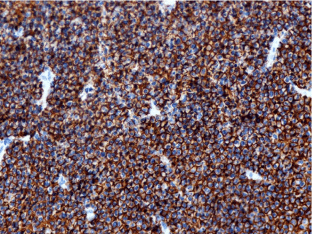
Figure 1
Diffuse intense membranous immunopositivity for CD20 indicating B lineage in a primary breast Marginal Zone Lymphoma. Immunohistochemistry
for CD20, X100.
Figure 2
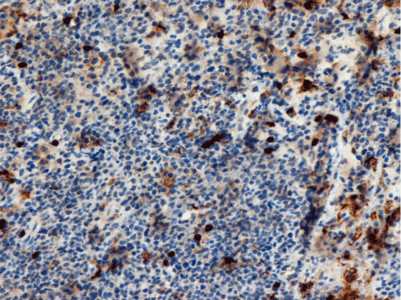
Figure 2
Primary breast Marginal Zone Lymphoma. Faint membranous
CD43 positivity in a small number of B cells, as opposed to the strong
staining of T cells. Immunohistochemistry for CD43, X100.
Figure 3
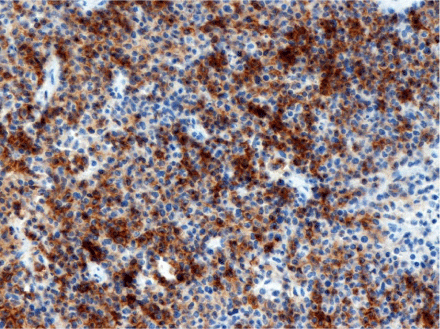
Figure 3
Primary breast Marginal Zone Lymphoma with B cell membranous
and cytoplasmic positivity for CD23. Immunohistochemistry for CD23, X100.
Discussion
The WHO classification distinguished three subtypes of MZL: Nodal, Splenic, and Extranodal
MZL (NMZL, SMZL and ENMZL) [8]. The latter defines a kind of
low grade mature B cell malignancies that arise mostly in the Mucosa-
Associated Lymphoid Tissue (MALT), whose most common site of
involvement is the gastrointestinal tract [7] or, less frequently, in nonmucosal
sites. Primary MZL of the breast is not a common entity [7].
It is important to distinguish ENMZL from other MZLs, as
well as from other subtypes of mature small B-cell lymphomas,
since these subsets present different clinical behaviors, treatment
and prognosis. In addition to NMZL, ENMZL and SMZL, small
B cell category includes Mantle Cell Lymphoma (MCL), Chronic
Lymphocytic Leukemia/Small Lymphocytic Lymphoma (CLL/SLL),
Lymphoplasmacytic Lymphoma (LPL), and Follicular Lymphoma
(FL).
Even though these lymphomas share some basic features that
make them join the same category, they are distinct entities with
several biologic and clinical differences [9]. The combination of
morphological and immune phenotypic evaluation can allow
the correct diagnosis and classification of these subsets [10].
Immunohistochemistry plays a key role in the differential diagnosis:
at present, with a panel of antibodies, most cases can be categorized
(Table 1). CD20 (or other B cell markers, such as CD19, PAX-5, CD79a
and CD79b) indicates B cell lineage, and is expressed on the great
majority of mature B-cell lymphomas. Other markers are useful in
differentiating a specific small B-cell lymphoid neoplasm from others,
i.e. CD5 and CD23 co-expression distinguishes CLL/SLL, and positive
nuclear staining of cyclin D1 is specific for MCL, as well as CD10 is
for FL [11-13]. With regard to the distinguishing immunophenotypic
pattern of MZL, CD20 shows a characteristic intense positivity, CD5
and CD10 are not expressed, CD23 is rarely positive (8%) and CD43
is variable (35%) [9,14,15]. Nowadays, also cytogenetics is a useful
tool for the diagnosis of some lymphomas, according to the presence
of a characteristic cytogenetic pattern in the specific disorder [16]. In
some Neoplasms, consistent cytogenetic abnormalities have not been
identified yet, while in others the identification of a specific pattern is
of main importance in the diagnosis, as it is the 11;14 translocation
for MCL [16]. With regard to MZLs, some recurrent chromosomal
translocation have been described. The most characterized of them
include t(11;18)(q21;q21), t(1;14)(p22;q32), and t(14;18)(q32;q21)
[17-19]. Trisomy 3 and trisomy 18 have also been reported [18,19].
Despite the advantage in classification, sometimes it is not easy
to make the definitive diagnosis ascribing a low grade lymphoma to
a precise subtype [20]. In the past, MZL and the extranodal variant
in particular were probably the most under-diagnosed mature B
cell neoplasms [11]. Still today several issues regarding low grade
lymphomas remain to be well-defined. The relative rarity of these
neoplasms is not helpful and the primary breast manifestation of a
lymphoma, especially MZL, is a rare entity [3-5,7].
Table 1

Table 1
Immunophenotype in the differential diagnosis of small B cell lymphomas.
Conclusion
With regard to breast tumors, not all lesions are of epithelial or
stromal origin, although these are the most common tumor types;
other kinds of neoplasms can occur on the breast, such as the ENMZL
of our case report. Treatment and prognosis may change according to
the neoplasm subtype, thus it is important to make the most precise
diagnosis as possible. The correct classification of different subsets
depends on the integration of clinical, morphological, cytogenetic
and, above all, immunohistochemical findings, as well as on the
pathologist's awareness that neoplasms can rarely arise in unusual
site. The rarity of some tumors requires the collaboration of clinicians
and pathologists in defining stringent diagnostic criteria; it would be
helpful for the pathologists to share their professional experiences.
We start with the report of a rare case. The idea of conducting
epidemiological surveys to design diagnostic criteria and clinical
features of rare neoplasms subtypes, is the challenge.
References
- Bray F, McCarron P, Parkin DM. The changing global patterns of female breast cancer incidence and mortality. Breast Cancer Res. 2004; 6: 229-239.
- Parkin DM, Bray F, Ferlay J, Pisani P. Estimating the world cancer burden: Globocan 2000. Int J Cancer. 2001; 94: 153-156.
- Martinelli G, Ryan G, Seymour JF, Nassi L, Steffanoni S, Alietti A, et al. Primary follicular and marginal-zone lymphoma of the breast: clinical features, prognostic factors and outcome: a study by the International Extranodal Lymphoma Study Group. Ann Oncol. 2009; 20: 1993-1999.
- Raghavan D, Blanke C, Johnson DH, Moots PL, Reaman GH, Rose PG, et al. 4th ed. Textbook of Uncommon Cancer. Hoboken: John Wiley & Sons; 2012. p225-277.
- Palazzo JP. Difficult Diagnoses in Breast Pathology. USA: Demos Medical Publishing; 2011; p196-242.
- Rock K, Rangaswamy G, O'Sullivan S, Coffey J. An Unusual Case of Marginal Zone B-Cell Lymphoma Arising in the Breast - Its Diagnosis and the Role of Radiotherapy in its Management. Breast Care (Basel). 2011; 6: 391-393.
- Kambouchner M, Godmer P, Guillevin L, Raphaël M, Droz D, Martin A. Low grade marginal zone B cell lymphoma of the breast associated with localised amyloidosis and corpora amylacea in a woman with long standing primary Sjögren's syndrome. J Clin Pathol. 2003; 56: 74-77.
- Swerdlow SH, Campo E, Harris NL, Jaffe ES, Pileri SA, Stein H, et al. WHO Classification of Tumours of Haematopoietic and Lymphoid Tissues. 4th ed. Lyon: IARC; 2008.
- His ED. Hematopathology: A Volume in the Series: Foundations in Diagnostic Pathology. 2nd ed. Elsevier; 2012. p203-384.
- Jaffe ES, Banks PM, Nathwani B, Said J, Swerdlow SH. Recommendations for the reporting of lymphoid neoplasms: a report from the Association of Directors of Anatomic and Surgical Pathology. The Ad Hoc Committee on reporting of lymphoid neoplasms. Hum Pathol. 2002; 33: 1064-1068.
- Rizzo K, Nassiri M. Diagnostic Workup of Small B Cell Lymphomas: A Laboratory Perspective. Lymphoma. 2012.
- Kilo MB, Dorfman DM. The utility of flow cytometric immunophenotypic analysis in the distinction of small lymphocytic lymphoma/chronic lymphocytic leukemia from mantle cell lymphoma. Am J Clin Pathol. 1996; 105: 451-457.
- Coelho Siqueira SA, Ferreira Alves VA, Beitler B, Otta MM, Nascimento Saldiva PH. Contribution of immunohistochemistry to small B-cell lymphoma classification. Appl Immunohistochem Mol Morphol. 2006; 14: 1-6.
- Extra nodal marginal zone lymphoma of breast and stomach. University Pathologists. 2011.
- Extranodal Marginal Zone B Cell Lymphoma. Differential Diagnosis, Stanford Medicine. 2006.
- Campbell LJ. Cytogenetics of lymphomas. Pathology. 2005; 37: 493-507.
- Armitage JO, Coiffier B, Dalla-Favera R, Harris NL. Non-Hodgkin Lymphomas. 2nd ed. Lippincott Williams & Wilkins; 2009; P232-251.
- Dierlamm J, Wlodarska I, Michaux L, Stefanova M, Hinz K, Van Den Berghe H, et al. Genetic abnormalities in marginal zone B-cell lymphoma. Hematol Oncol. 2000; 18: 1-13.
- Cuneo A, Castoldi GL. Marginal zone B-cell lymphoma. Atlas Genet Cytogenet Oncol Haematol. 2006; 10: 180-183.
- Thieblemont C. Clinical presentation and management of marginal zone lymphomas. Hematology Am Soc Hematol Educ Program. 2005; 307-313.