Clinical Image
Bronchial Malt Lymphoma Successfully Treated by Helicobacter Eradication
Kota Murohashi, Yu Hara*, Takashi Sato and Takeshi Kaneko
Department of Pulmonology, Yokohama City University Graduate School of Medicine, Japan
*Corresponding author: Yu Hara, Department of Pulmonology, Yokohama City University Graduate School of Medicine, 4-57 Fukuura, Kanazawa-ku, Yokohama City, 236- 0024, Japan
Published: 16 May, 2018
Cite this article as: Murohashi K, Hara Y, Sato T, Kaneko T.
Bronchial Malt Lymphoma Successfully
Treated by Helicobacter Eradication.
Clin Oncol. 2018; 3: 1463.
Keywords
Primary bronchial MALT lymphoma; Clarithromycin; Helicobacter eradication
Clinical Image
A 79-year-old woman was admitted to our hospital due to a finding of left main bronchial wall irregularity on chest CT. The bronchoscopic findings revealed a number of white, smooth, glossy granules on the left main bronchus (Figure 1A). Histological findings of bronchial biopsy specimens demonstrated outgrowths of diffuse lymphoid cells and lymphoepithelial lesions (Figure 2A). Immuno-histochemical staining revealed positive for CD20 and CD79a, and negative results for CD5, CD10, and CD43 (Figure 2B). In addition, both gastroscopy and colonoscopy were performed, but revealed no evidence of Mucosa-Associated Lymphoid Tissue (MALT) lymphoma. Coincidentally, the bronchial wall lesions were improved by 3 months after successful Helicobacter eradication (using clarithromycin, amoxicillin and proton pump inhibitors) as confirmed by serological testing (Figure 1B). The effect of Helicobacter eradication on primary bronchial MALT lymphoma is unclear [1], but several case reports have suggested that clarithromycin was effective in the treatment of primary bronchial MALT lymphoma [2].
Figure 1
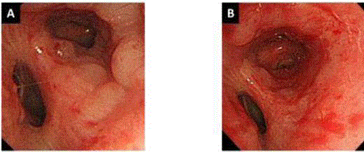
Figure 1
A) Bronchoscopic findings on admission reveal a number of smooth, white, glossy granules on the
left main bronchus. B) Bronchoscopic findings after Helicobacter eradication reveal improvement of bronchial
wall lesions.
Figure 2
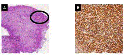
Figure 2
A) Histological examination of a bronchial biopsy specimen demonstrates outgrowths of diffuse
lymphoid cells and lymphoepithelial lesions (black circle). Helicobacter species is not evident. B) Immunohistochemical
stain showing positive results for CD20.
References
- Grünberger B, Wöhrer S, Streubel B, Formanek M, Petkov V, Puespoek A, et al. Antibiotic treatment is not effective in patients infected with Helicobacter pylori suffering from extragastric MALT lymphoma. J Clin Oncol. 2006;24(9):1370-5.
- Ishimatsu Y, Mukae H, Matsumoto K, Harada T, Hara A, Hara S, et al. Two cases with pulmonary mucosa-associated lymphoid tissue lymphoma successfully treated with clarithromycin. Chest. 2010;138(3):730-3.