Clinical Image
Unusual Endoscopic Findings of Intestinal Spirochetosis
Jun Nishikawa1*, Osamu Miura2, Shinichi Hashimoto3 and Isao Sakaida3
1Department of Laboratory Science, Yamaguchi University Graduate School of Medicine, Japan
2Department of Gastroenterology, Hofu Institute of Gastroenterology, Japan
3Department of Gastroenterology and Hepatology, Yamaguchi University Graduate School of Medicine, Japan
*Corresponding author: Jun Nishikawa, Department of Laboratory Science, Yamaguchi University Graduate School of Medicine, 1-1-1 Minamikogushi, Ube, Yamaguchi 755-8505, Japan
Published: 25 Mar, 2018
Cite this article as: Nishikawa J, Miura O, Hashimoto
S, Sakaida I. Unusual Endoscopic
Findings of Intestinal Spirochetosis. Clin
Oncol. 2018; 3: 1451.
Keywords
Intestinal Spirochetosis; White-coated Elevation; Pseudo Brush Border
Clinical Image
We performed a screening colonoscopy on a 50-year old man who was scheduledto undergo right hepatectomy for hepatocellular carcinoma with hepatitis C virus infection. He did not have any symptoms such as diarrhea or blood test abnormalities. Polypoid lesions with white friable mucosa were detected in the transverse colon. The lesions were segmented, and unaffected mucosa was present between them (Figure 1 and 2). Histologic examination of a biopsy specimen from the lesions showed a fuzzy fringe covering the colonic mucosa called a pseudo-brush border. Immunohistochemistry using anti-Treponema pallidum antibody revealed positive staining of the surface covering the epithelial cells (Figure 3 and 4). These findings were consistent with intestinal spirochetosis. We administered 1000mg metronidazole for 7 days. Follow-up colonoscopy showed that the multiple lesions in the transverse colon had disappeared. Intestinal spirochetosis can be accidentally diagnosed by mucosal biopsy because there are no remarkable endoscopic findings [1]. We propose that whitecoated elevations found segmentally in the colon may be one of the endoscopic findings of intestinal spirochetosis.
Figure 1
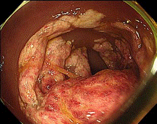
Figure 1
Figure 2
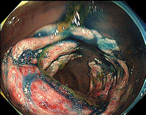
Figure 2
Figure 3
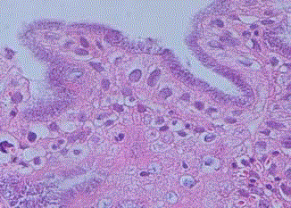
Figure 3
Figure 4
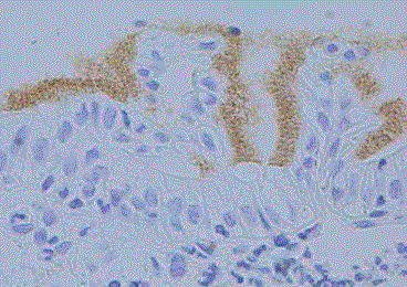
Figure 4