Clinical Image
Deciphering the Difficulty for Pathologic Diagnosis of Hodgkin Lymphomas
Ke-seng Zhao and Meigang Zhu*
Department of Pathology and Pathophysiology, Southern Medical University Guangzhou, China
*Corresponding author: Ke-seng Zhao, Department of Pathophysiology, Southern Medical University, China
Published: 19 Apr, 2018
Cite this article as: Ke-seng Zhao, Zhu M. Deciphering the
Difficulty for Pathologic Diagnosis of
Hodgkin Lymphomas. Clin Oncol. 2018;
3: 1444.
Clinical Image
Hodgkin Lymphoma (HL) is composed of 30% of lymphoma. It is difficult for pathologic
diagnosis because of a variety of morphology in Reed- Sternberg Cells (R-S cell) and reactive
inflammatory cells on the background [1-3]. According to our experience, following points may
help the diagnosis of HL:
1. The size of diagnostic R-S cell is 2 times than one B-immunoblast or one histocyte in the
same field of section (Figure 1) [4].
2. The diagnostic R-S cell usually has 2 nuclei, which includes big nucleolus. The size of
nucleolus in diagnostic R-S cell is equivalent to the size of one
erythrocyte or one small lymphocyte (about 5 μm) in the same field
of section (Figure 2) [4].
3. The appearances of series R-S cells well help to diagnose
HL. A series R-S cells includes more than 2 types of R-S cells, i.e. one
is diagnostic R-S cells ( with 2 nuclei ) served as a marker, others are
one or more variant R-S cells, including mononuclei, multinuclei,
lacuner, mummigied R-S cells (Figure 3) [4].
4. Quantitative indicator of R-S cell helps for classified
diagnosis of CHL. i.e. 5-15 R-S cells/HPF for Mixed Cellular CHL
(MCCHL), >15 cells/HPF for Lymphocyte Depleted CHL (LDCHL)
(Figure 4). <5 cells/HPF for Lymphocyte Rich CHL (LRCHL) [2,5].
Figure 1
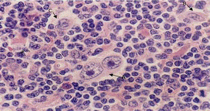
Figure 1
The size of a diagnostic R-S cell A) Is 2 times than a histocyte. B) A B-immunoblast. C) In the same
field.
Figure 2
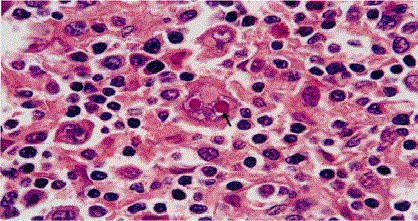
Figure 2
The size of diagnostic R-S cell nucleolus (arrow) is equivalent to the size of small lymphocytes around
the R-S cell.
Figure 3
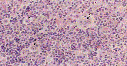
Figure 3
Series R-S cells include A-Diagnostic R-S cells (two nuclei), B-variant R-S cell with mononucleus;
C-variant R-S cell with multi- nuclei.
Figure 4
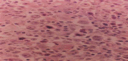
Figure 4
The amount of series R-S cells is more than 15 cells/HPF in
LDCHL.
References
- Swerdlow SH, Campo E, Harris NL, Jaffe ES, Pileri SA, Stein H, et al. WHO Classification of Tumors of Hematopoietic and Lymphoid Tissues. Lyon. 2017;2:423-41.
- Jaffe ES, Harris NL, Vadiman JW, et al. Hematopathology Classical Hodgkin lymphoma. Elsevier. 2013:454.
- Fletcher CM. Diagnostic Histopathology of Tumors: volume 2. Tumors of lynyshoreticular system. 3rd ed. Elsevier. 2007:1139.
- Zhu MG, Huang J. Differential Diagnosis between Benign and Malignant of Lymphoid Issues Proliferative Lesions. Guangdong Science & Technology Press. 2012;314-33.
- Lochim HL, Mecteirros LJ. Lochim’s Lymph Node Pathology, Lippincott Williams and Wilkins. 2008:306.