Review Article
Modified Transpectoral Approach for Ultrasound Guided Axillary Lymph Node Core Biopsy in Challenging Cases
Pascaline S*, Black D, Woodland K and Yaroshenko O
Department of Breast Radiologist, Kettering General Hospital, UK
*Corresponding author: Pascaline S, Department of Breast Radiologist, Kettering General Hospital, UK
Published: 06 Jan, 2018
Cite this article as: Pascaline S, Black D, Woodland K,
Yaroshenko O. Modified Transpectoral
Approach for Ultrasound Guided
Axillary Lymph Node Core Biopsy in
Challenging Cases. Clin Oncol. 2018;
3: 1407.
Abstract
The axillary nodal status is a very important factor in the preoperative staging in breast cancer patients. Our department for the last three years has utilised predominantly US guided core biopsy 14G, and in cases with limited access or anatomical difficulties we reverted to utilising FNA sampling. The standard widely used approach for these procedures is from the infero-lateral to supero-medial aspect towards the target to avoid major vessels and muscles. To optimise access to the axilla, a wedge pillow is placed under the patient’s back to rotate the patient’s body and elevate the targeted area.
Introduction
Occasionally axillary biopsy has to be performed for patients with significantly reduced mobility
of the shoulder, or patients with very high or extremely low BMI (1-7). The axillary lymph nodes
in these cases still could be scanned but biopsy due to reduced anatomical access is technically
challenging. Lymph nodes may be too deep or positioned just above vessels. Concave or excessively
full axilla often reduces the possibility of a safe approach (Figure 1 and 2).
The modified method described below was once developed in our breast clinic for a particular
patient with a healing shoulder fracture and since then used in complex cases when standard
approach is not feasible to sample suspicious lymph nodes (2,5).
Discussion
The axillary nodal status is a very important factor in pre-operative staging in breast cancer
patients. It is crucial in pre-op planning between a sentinel node biopsy oran axillary lymph node
dissection.
As described above, this inventive core biopsy method of sampling non-palpable axillary
lymph nodes is performed using a modified reverse transpectoral approach. 5 ml of 2 % Xylocaine
isinjected for superficial and deep local anesthesia.Fourteen-gauge (2.1 mm) needle was used with
an automatic biopsy gun, and 22 mm notchselected (Bard-Magnum Biopsy Instrument) with three
needle passes usually.
I am aware that transpectoral approach can be more painful therefore few minutes longerpause
between local anesthetics injection and biopsyused consciously to
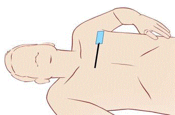
achieve better anesthesia (Figure 3 and 4). 16-18G needles or halfnotch
sampling can be used according to the patient build and lymph
node position. To our best knowledge, previous studies performed
the core biopsy by using 16-gauge or 18-gauge needles (Figure 5), and
there are a few reports about the 14-gauge needle core biopsy of the
axilla lymph node [7-10].
A special care should be taken for reducing unwanted
complications by fully acknowledging detailed anatomy including
vessels and nerves in the axilla. Proper care andan individual approach
should be applied while positioning the patient and performing
ultrasound guided biopsy (Figure 6), both with the traditional
approach as well as with described above modified approach, to avoid
traumatic injuries during the large needle core biopsy of the axilla.
In conclusion modified transpectoral ultrasound guided core
biopsyof the axillary lymph nodes could be an effective alternative
to surgical biopsy for the histological diagnosis in certain complex
challenging cases.
Figure 1
Figure 1
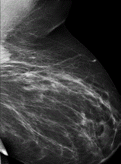
46 yoHigh BMI patient with reduced left shoulder mobility. She presented symptomatically with large
axillary tail malignant looking mass visible on MLO view only.
Figure 2
Figure 2
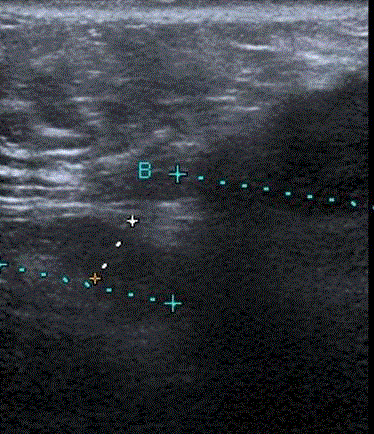
Conventional axillary US demonstrates a large malignant irregular
hypoechoic mass and adjacent to it craniomedially an abnormal looking
lymph node. Only the mass was biopsied at that occasion.
Figure 3
Figure 3
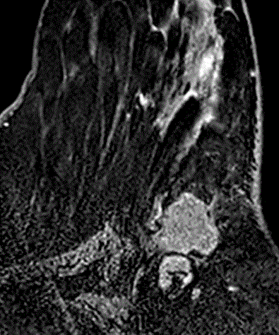
Breast MRI confirming challenging proximity of the biopsy proven
cancerand abnormal looking lymph node.
Figure 4
Figure 4
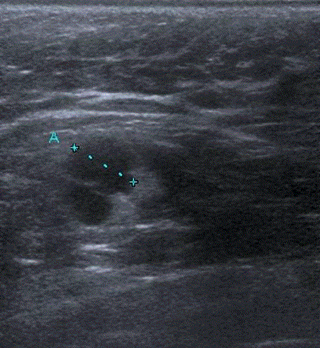
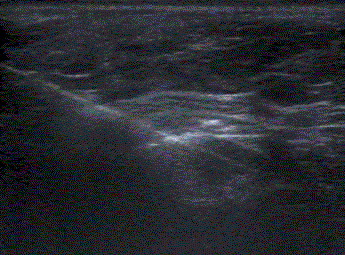
Transpectoral US of the area of interest.Using the modified
ultrasound technique the abnormal lymphnode is now closer to the transducer
and there is no anatomical limitation of the shoulder, underlying vessels or
overlying breast cancer.
Figure 5
Figure 5
Modified position for the scan on previous image and subsequent
US guided core biopsy. The maximal stretching and flattening of the pectoral
muscle and overlying axillary soft tissue is noted.
Figure 6
Figure 6
Transpectoral biopsy with reverse approach – from superomedial to
lateral, parallel to the chest wall, enables a safer sampling of the challenging
lymph node. Metastatic deposits were confirmed on histopathology.
References
- Topal U, Punar S, Taşdelen I, Adim SB. “Role of ultrasound-guided core needle biopsy of axillary lymph nodes in the initial staging of breast carcinoma,” European Journal of Radiology, vol. 2005; 56: 382-385.
- H Abe, R A Schmidt, C A Sennett, A Shimauchi, G M Newstead, “USguided core needle biopsy of axillary lymph nodes in patients with breast cancer: why and how to do it,” Radiographics, vol. 2007; S91–S99, 2007.
- Houssami N, Ciatto S, Turner RM, Cody HS 3rd, Macaskill P. MacAskill, Preoperative ultrasound-guided needle biopsy of axillary nodes in invasive breast cancer: meta-analysis of its accuracy and utility in staging the axilla. Annals of Surgery, vol. 2011; 254: 243-251.
- Rautiainen S, Masarwah A, Sudah M, Sutela A, Pelkonen O, Joukainen S, et al. Axillary lymph node biopsy in newly diagnosed breast cancer: comparative accuracy of fine-needle aspiration biopsy versus core needle biopsy. Radiology, vol. 2013; 269:54-60.
- Lernevall A. Imaging of axillary lymph nodes. ActaOncol. 2000; 39: 277- 281.
- deKanter AY, van Eijck CH, van Geel AN, Kruijt RH, Henzen SC, Paul MA, et al. Multicentre study of ultrasonographically guided axillary node biopsy in patients with breast cancer. Br J Surg. 1999; 86:1459-1462.
- Ki Hong Kim. The Safety and Efficiency of the Ultrasound-guided Large Needle Core Biopsy of Axilla Lymph Nodes. Yonsei Med J. 2008; 49(2): 249-254.
- Damera A, Evans AJ, Cornford EJ, Wilson AR, Burrell HC, James JJ, et al. Diagnosis of axillary nodal metastases by ultrasound-guided core biopsy in primary operable breast cancer. Br J Cancer. 2003; 89:1310-1313.
- Susini T, Nori J, Vanzi E, Livi L, Pecchioni S, Bazzocchi M, et al. Axillary ultrasound scanning in the follow-up of breast cancer patients undergoing sentinel node biopsy. Breast.2007;16:190-196.
- Nori J, Bazzocchi M, Boeri C, Vanzi E, Nori Bufalini F, Mangialavori G, et al. Role of axillary lymph node ultrasound and large core biopsy in the preoperative assessment of patients selected for sentinel node biopsy. Radiol Med (Torino) 2005;109:330.

