Mini Review
Clinical Grand Rounds in Pediatric Oncology
Suhasini Gazula*, Ms Ruby R, Leela rani V and Rajasekhar A
Departments of Pediatric Surgery, Employees State Insurance Corporation (ESIC) Medical College, India
*Corresponding author: Suhasini Gazula, Department of Pediatric Surgery, Employees State Insurance Corporation (ESIC) Medical College, India
Published: 06 Jan, 2018
Cite this article as: Gazula S, Ms Ruby R, Leela rani V,
Rajasekhar A. Clinical Grand Rounds in
Pediatric Oncology. Clin Oncol. 2018;
3: 1403.
Case
An 8-day-old female neonate presented with antenatally detected mass in the right kidney.
On Examination
Per abdomen: hard mass with well defined margins palpable in the right hypochondrium and right lumbar region, approximately 6 cm × 10 cm.
Investigations
Usg-abdomen and pelvis with Doppler
Well defined large heterogenous solid lesion of size 5.7 cm × 5.6 cm seen in right kidney medially
displacing the Inferior vena cava. IVC upper segment seen but rest of IVC and right renal vein can’t
be visualised- probable compression by the mass. Left kidney, liver normal.
CECT- Abdomen: Well-defined heterogeneous enhancing solid mass lesion seen arising from
the upper and mid poles of right kidney? Tumor.
Usg guided fnac: Features of Benign spindle cell lesion.
Major surgical profile
PET Scan (Post-operative): No residual tumor, No distant metastasis.
Pre-op management
• Baby admitted in ICU, maintained thermoregulation and vitals monitored
• Administered pre-operative vitamin K and antibiotics and shifted in transport incubator
to the operating room for an exploratory laparotomy + precede right nephroureterectomy.
Operative findings
• Right Renal tumour ~8 cm × 5 cm; No enlarged lymph nodes seen
• Ureter identified and mobilized till the pelvic part and ureterectomy done
• Renal vein and artery identified and transfixed
• Right Nephrectomy done preserving right adrenal gland
• Tumor bed margins biopsied and marked with clips
Post-operative management
• To 1st Post-Operative Day (POD)
• Baby was kept in Pediatric Surgery ICU and Nil per Oral
• IV antibiotics & fluids, vitals and abdominal girth charting
• IV Fentanyl infusion for pain relief
• On 2nd POD - Started Breast feeds
• On 5th POD- Child shifted to ward
• On 7th POD-Child discharged from hospital with uneventful recovery
Histopathology Findings
• Well circumscribed tumour tissue with infiltrating margins and composed of cellular
growth of spindle cells and interlacing fascicles with papillary , acidophilic cytoplasm and bland
spindled nuclei
• There are entrapped glomeruli and tubules in the tumour
tissue
• Focal areas shows cellular proliferation with increased
mitotic activity
• There is no invasion in to renal pelvis or post renal fat or
lymphovascular invasion
• All margins are free from tumour
• Based on operative and histopathology findings it was
classified as right congenital mixed mesoblastic nephroma (classical
+ cellular)
Figures
Figures
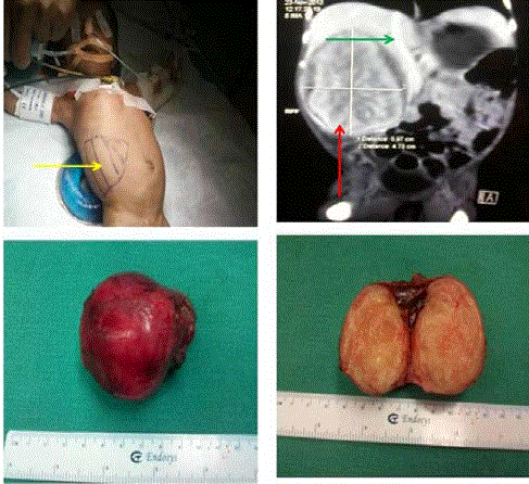
1). 8 cm x 5 cm right renal tumor 2). CECT abdomen showing
large renal tumor (red arrow) pushing IVC (green arrow) medially 3).
Tumour resected into to 4). Cut-section of tumor having uterine fibroid-like
appearance.
Adjuvant Chemotherapy
In lieu of mixed variant on histopathology with potential for
recurrence, PET scan was done which ruled out any other distant
metastasis and baby underwent 8 weeks of Vincristine + Actinomycin-
D chemotherapy in our ward which she tolerated very well.
Present status
Baby is 5 years old now, healthy and disease free.
Congenital Mesoblastic Nephroma
Congenital Mesoblastic nephroma is a rare neonatal/pediatric
renal tumor that is usually found before birth by USG or within the
first 3 month of life. It is similar in gross and histologic appearance
to a uterine leiomyoma with spindled cell bundles, but composed of
immature renal stromal cells. The tumor lacks renal blastema and
neoplastic metanephric elements, thereby differentiating it from
Wilm’s tumor. It can be detected antenatally especially with judicious
use of ultrasonography. There are two pathologic variants: classic
CMN and atypical or cellular CMN. The classic form is characterized
by rare mitoses and absence of necrosis. Atypical or cellular CMN
is characterized by a high mitotic index, hypercellularity, necrosis,
hemorrhage, and invasion of adjacent structures warranting use of
adjuvant chemotherapy.
Therapeutic management
Upfront surgical excision+Chemotherapy+Radiotherapy
Points to Remember
• Cancer can occur in children – even in newborn babies!!
• Cancers in babies can sometimes be detected even before
birth by antenatal ultrasonography
• Cancers in children are treatable and when detected early
have very good outcomes
• Paediatric cancers do not cause associated symptoms
like weight loss, weakness except in late stages - so do not wait for
symptoms
Do not waste time & seek urgent pediatric specialist care.

