Short Communication
Experience of Early Gastric Cancer at Dual Energy CT with Iodine Map
Manabu Watanabe*
Department of Gastroenterology and Hepatology, Toho University Medical Center, Ohashi Hospital Address: 2-17-6 Ohashi Megro-ku Tokyo 153-8515, Japan
*Corresponding author: Manabu Watanabe, Department of Gastroenterology and Hepatology, Toho University Medical Center, Ohashi Hospital, 2-17-6 Ohashi Megro-ku, Tokyo 153-8515, Japan
Published: 31 Dec, 2017
Cite this article as: Watanabe M. Novel RNA Editing Sites
of 5-HT2C Receptor Encoding the
Third Intracellular Loop Domain in
Undifferentiated NG108-15 Cells. Clin
Oncol. 2017; 2: 1383.
Short Communication
The patient was a 77-year-old man in whom posterior wall thickening of the lower gastric body
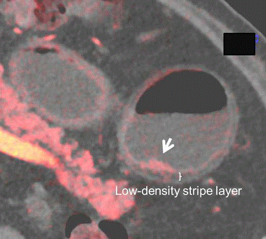
was shown in abdominal CT. In the arterial phase of iodine map obtained with dual energy CT
(DECT), the lesion showed the mucosal enhancement compared to the adjacent mucosal layer
(Figure 1). There was no enhancement in the submucosal low-density-stripe layer, including in
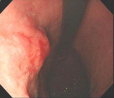
the portal and equilibrium phases. The type 0-IIa lesion that bled easily was found by gastroscopy
(Figure 2) and was diagnosed as a well-differentiated adenocarcinoma (tub1/pap, SM1). DECT is
based on the simultaneous acquisition of two data sets of the body area under study using different
voltage (80 kVp and 140 kVp) [1,2]. At low-energy (80 kVp), the contrast of the agent is strong
despite a high noise level, while at high-energy (140kVp) the contrast and noise level are low. With
a tube voltage of 120 kVp, which is equivalent to the synthesized voltage of these two levels, images
with characteristics of both voltages can be obtained. Furthermore, iodine map, which is one of
DECT applications, can be superimposed on morphologic imaging to show contrast enhancement.
Among the types of gastric cancer, well-differentiated adenocarcinoma has developed atypical
ducts and a large number of capillaries, and thus well-enhancing mucosal thickening can be detected
by dynamic CT in the early phase. In contrast, poorly-differentiated adenocarcinoma is characterized
by a desquamated epithelium due to ducts with poor differentiation or development of signet ring
cells in the interstitium, and thus mucosal enhancement is not observed in the early phase. Based
on these pathological characteristics, the invasion depth of gastric cancer has been diagnosed by
MDCT in some cases, but the accuracy is still debatable3. However,
DECT may be useful both for diagnosis of gastric cancer and other
gastrointestinal cancers, and for accurate evaluation of the invasion
depth.
Figure 1
Figure 1
The iodine map obtained with dual energy CT in the arterial phase showed well-enhancing mucosal
thickening with intact low-density stripe layer at the posterior wall of the lower gastric body (arrow).
Figure 2
References
- Zhang LJ, Peng J, Wu SY, Wang ZJ, Wu XS, Zhou CS, et, al. Liver virtual non-enhanced CT with dual-source, dual-energy CT: a preliminary study. Eur Radiol. 2010; 20: 2257-64.
- Heye T, Nelson RC, Ho LM, Marin D, Boll DT. Dual-energy CT applications in the abdomen. AJR Am J Roentgenol. 2012;199: S64-70.
- Kim JW, Shin SS, Heo SH, Choi YD, Lim HS, Park YK, et al. Diagnostic performance of 64-section CT using CT gastrography in preoperative T staging of gastric cancer according to 7th edition of AJCC cancer staging manual. Eur Radiol. 2012; 22:654-62.


