Case Report
Organizing Pneumonia in a Patient with Myelodysplastic Syndrome Who was Treated with Azacitidine
Masahiro Manabe1*, Gakuya Tamagaki2, Yuji Hagiwara3, Reiko Asada3, Dai Momose1, Yasuyoshi Sugano1, Yuko Kuwae4 and Ki-Ryang Koh1
1Department of Hematology, Osaka General Hospital of West Japan Railway Company, Japan
2Department of Respiratory Medicine, Osaka General Hospital of West Japan Railway Company, Japan
3Department of Clinical Laboratory, Osaka General Hospital of West Japan Railway Company, Japan
4Department of Pathology, Graduate School of Medicine, Osaka City University, Japan
*Corresponding author: Masahiro Manabe, Department of Hematology, Osaka General Hospital of West Japan Railway Company, Japan
Published: 03 Dec, 2017
Cite this article as: Manabe M, Tamagaki G, Hagiwara Y,
Asada R, Momose D, Sugano Y, et al.
Organizing Pneumonia in a Patient with
Myelodysplastic Syndrome Who was
Treated with Azacitidine. Clin Oncol.
2017; 2: 1378.
Case Presentation
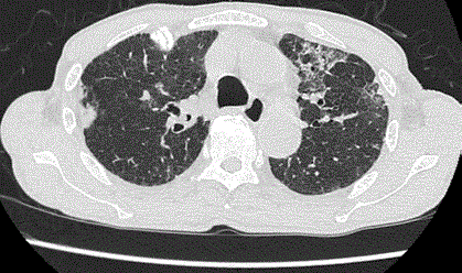
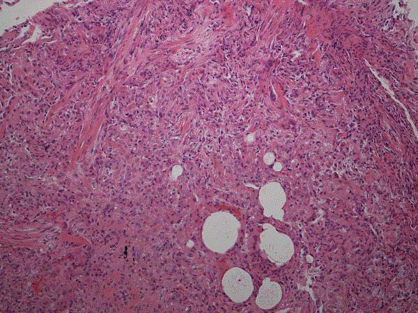
A 75-year-old male was referred to our hospital due to anemia. His blood data showed a hemoglobin concentration of 5.7g/dL, and a white blood cell count of 2300/μL (blasts: 16.5%). A bone marrow aspiration showed trilineage dysplasia and a blast count of 13.0%. The patient was diagnosed with myelodysplastic syndrome with excess blasts-2. He was started on azacitidine monotherapy. Three weeks after the initiation of the third course of treatment, he presented with a fever, dry cough, and dyspnea on effort. Computed tomography revealed bilateral interstitial infiltrates (Figure 1), and spirometry tests demonstrated a markedly restrictive pattern. He underwent a transbronchial lung biopsy, which revealed organizing lesions (Figure 2). A pathological diagnosis of organizing pneumonia was made. Although it seems to be a very rare complication, it is important for hematologists to be aware that interstitial lung disease can occur as an adverse event of azacitidine use.
Figure 1
Figure 1
At the time that the patient presented with respiratory symptoms, CT revealed bilateral abnormal
shadows, which were compatible with OP.


