Clinical Image
Early Pharyngeal Cancer Treated by ESD
Koichiro Kawaguchi, Hiroki Kurumi and Hajime Isomoto*
Department of Gastroenterology, Tottori University Hospital, Japan
*Corresponding author: Hajime Isomoto, Department of Gastroenterology, Tottori University Hospital, Japan
Published: 10 Nov, 2017
Cite this article as: Kawaguchi K, Kurumi H, Isomoto H.
Early Pharyngeal Cancer Treated by
ESD. Clin Oncol. 2017; 2: 1363.
Clinical Image
In recent years, improvement of image quality of endoscopes, especially progress of image
enhanced endoscopy (IEE) has made it possible to point out neoplastic lesions in pharyngeal
laryngeal region even in the early stage byendoscopists. In addition, classification of differential
diagnosis and depth of reach diagnosis of neoplastic lesions has been established by the evaluation
of intra-papillary capillary loop by the magnified endoscope with IEE, and these lesions can be
accurately evaluated.
Furthermore, due to advances in endoscopic treatment typified by endoscopic submucosal
dissection (ESD), lesions that can be cured with endoscopic treatment alone have come into
this area. Endoscopic treatment was not only minimally invasive for patients, it also great merit
of function preservation. It is no exaggeration to say that progress in endoscopic technology has
changed the concept of diagnosis and treatment of neoplastic lesions in the field of otolaryngology.
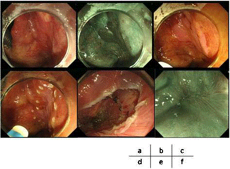
Figure shows our case to whom ESD was successfully performed.
Figure Legend
a: a slightly reddish mucosa with standard pharyngeal endoscopy
b: image-enhanced endscopic (IEE) view as a brownish area consisting of abnormal intrapapillary
capillary loops and was confirmed iodine-unstained lesion (c).
d: The lesion was marked and endoscopic submucosal dissection was successfully accomplished
en bloc (e).
e: Follow-up IEE showed that here has been no recurrence with only scar preserving swallow
function.
Figure
Figure
(Case 1, diagnosis): Case of 0-IIb type early cancer (EP) presentin right hypo-pharyngeal region, but
white light imaging was difficult to detect (a). NBI could detect the lesion as the “brownish area” (b). Magnified
endoscopic imaging showed enlarged IPCL(c-e). The patient’s esophagus showed “multiple Lugol-void lesion”
(f).
(Case 1, treatment): Observation of lesions under intubated general anesthesia, white light (a), NBI (b), Lugor
staining (c). Marking around the lesion with Dual knife (d). The lesion was resected en-blocby ESD (e).After six
months, the ulcer was completely closed and there was no recurrence (f).
Case 2: Case of 0-IIb+IIa type early cancer (EP)presentin left hypo-pharyngeal region, white light (a), NBI (b).
Submucosal dissection was performed with traction by the otologist (c). After ESD, hypopharynx was swelling
(d), and the muscular layer was exposed to the ulcer bed (e). The lesion was resected en-blocby ESD(f).

