Case Report
Acute Esophageal Necrosis Complicated by Severe Upper Gastrointestinal Bleeding in Association with Bosutinib Treatment for Chronic Myelogenous Leukemia
Danah Mohammad1, Anwar Albasri1, Jeff Lipton2* and Michael Beyak1
1Department of Medicine, Queens University, Kingston, ON, Canada
2Department of Medical Oncology and Hematology, University of Toronto, Toronto, Canada
*Corresponding author: Jeff Lipton, Department of Medical Oncology and Hematology, Princess Margaret Cancer Centre, University of Toronto, Toronto, Ontario, Canada
Published: 26 Apr, 2017
Cite this article as: Mohammad D, Albasri A, Lipton J,
Beyak M. Acute Esophageal Necrosis
Complicated by Severe Upper
Gastrointestinal Bleeding in Association
with Bosutinib Treatment for Chronic
Myelogenous Leukemia. Clin Oncol.
2017; 2: 1266.
Abstract
Acute Esophageal Necrosis (AEN), also known as “Black Esophagus”, is a rare life threatening condition with unknown cause. We present a case of AEN in an 82 years-old male on Tyrosine Kinase Inhibitor (TKI) for Chronic Myelogenous Leukemia (CML). While TKI is known to cause GI toxicity, this is the first case, to our knowledge, reporting AEN.
Introduction
AEN is an uncommon clinical entity caused by variety of factors. It was first described by
Goldenberg et al in 1990 [1]. The diagnosis is based on endoscopic appearance after excluding
ingestion of corrosive agents. In most patients, the resolution of endoscopic findings occurs with
supportive care [2]. Mortality is high and it is largely due to the underlying disease rather than being
directly attributable to complications of acute esophageal necrosis.
We report a case of AEN in a patient with CML. Although the main cause of AEN is unknown,
our case was temporally associated with treatment of CML with a TKI, bosutinib. At first it was
thought that the effect of TKI at the molecular mechanism level appears to comprise only a targeted
approach in blocking tyrosine kinases. However, this should not be misleading as many closely
interconnected signaling pathways are involved. TKIs are a new modality of anti-cancer-therapy
amending classical cytotoxic regimes. TKIs are of substantial benefit in terms of efficacy with a
tolerable safety profile. However, long-term safety issues might not be fully elucidated at present
and, thus, cannot be finally judged upon.
Bosutinib is an oral TKI for a philadelphia chromosome positive (Ph+) CML. In general, TKIs
are associated with gastrointestinal toxicity, however, bosutinib has higher degree of gastrointestinal
adverse effects. Bosutinib gastrointestinal adverse effect profile included diarrhea, vomiting, nausea,
and abdominal pain [3]. Currently, available studies have not specifically reported on esophageal
adverse effect associated with bosutinib treatment.
Case Report
An 82-year-old male with a history of chronic phase CML presented with four days’ of
hematemesis and melena followed by an episode of syncope. The TKA, Bosutinib 300mg oral daily,
had been started two weeks prior to presentation. In addition, he had a past medical history of
hypertension, and gastroesophageal reflux disease.
On physical examination, he was found to be hypotensive with blood pressure of 90/55mmHg
and tachycardiac with heart rate of 110 beats per minute. Three black tarry loose bowel movements
were witnessed in the emergency room. He was resuscitated with Intravenous crystalloid and started
on intravenous PPI. Although the blood pressure has dropped in our patient, we opt to follow the
restrictive transfusion strategy with a transfusion threshold of 70g/L. The restrictive transfusion
has better outcome in patients with upper gastrointestinal bleeding. This strategy reduces the risk
of further bleeding, the need for rescue therapy, and the complication rate, all while improving the
survival rate [4].
Laboratory investigations revealed initial hemoglobin of 141 g/L, falling to a nadir of 128 g/L.
There was evidence of acute kidney injury with urea of 22.8 mmol/L and creatinine of 185 umol/L.
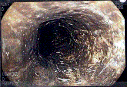
Upper endoscopy was performed within 2 hours of arrival at our hospital and revealed a confluent black esophageal mucosa, consistent with mucosal necrosis, with
a distinct transition to normal mucosa at the gastroesophageal
junction. In addition, a non-bleeding visible vessel was seen just
proximal to the gastroesophageal junction was seen. This was treated
with application of a single clip.
The AEN was managed conservatively with oral PPI,
discontinuation of bosutinib and advancing the diet as tolerated. He
was discharged from hospital four days later. However, four days later
he presented again with a massive upper GI bleed, with hematemesis,
melena and hematochezia, complicated by significant hypotension
requiring ICU admission. Upper endoscopy was repeated, showing
resolving esophageal necrosis, but an active bleeding visible vessel
again just above the gastroesophageal junction. Hemostasis was
achieved after injection of epinephrine (1:10,000 concentration) and
placement of another clip.
Figure 1
Discussion
AEN, also known as “Black Esophagus”, is a rare life threatening
condition with an incidence ranging between 0.01 to 0.28% [4]. It
affects males more than females in a 4:1 ratio, with a peak incidence
in the 6th decade of life [2]. Mortality ranges from 15% up to 36% with
only less than 10% of cases have been reported as a cause of death due
to esophageal disease itself [5-8].
The exact etiology is unknown but it is thought to be a result of
multiple pathological insults. These include local ischemia, corrosive
acid exposure and decreased esophageal clearance. Risk factors
that identified in the literature include, but not limited to, shock,
hypoperfusion, infection, hyperglycemia, underlying malignancy,
and irradiation.
The most common presentation of AEN is upper GI bleeding.
Other manifestations include retrosternal discomfort, epigastric pain
and dysphagia [9,10]. It can be complicated by esophageal perforation
in less than 7% while esophageal strictures may occur in more than
10% of patients. The black discoloration with a sharp transition to
normal mucosa of the gastroesophageal junction is the hallmark of
diagnosing AEN endoscopically. Although biopsy is needed to rule
out other causes of black esophagus, the resolution of the endoscopic
finding on repeated scope confirmed the diagnosis of AEN in our
patient.
Similar to reported cases in the literature, the exact etiology of
AEN is unknown in our patient. Hypotension was common found
amongst those with AEN. It has been hypothesized that acute circulatory compromise may contribute to the esophageal ischemic
necrosis. However, it is difficult to differentiate whether it is the cause,
or the effect from the bleeding. We propose the two-hit hypothesis
whereby bosutinib, the first “hit”, predisposes the esophageal mucosa
to ischemic compromise via an unknown mechanism. Thereafter, the
second “hit”, acute hypotension, precipitates the esophageal necrosis.
Given the recent start of the TKI, bosutinib, and the absence of other
significant potential causes, it seemed reasonable to implicate the drug
in this case of AEN. The safety profile of bosutinib was assessed and it
was found that gastrointestinal toxicity is more common in boustinib
compared to other tyrosine kinases inhibitors. Gastrointestinal
adverse effects that were associated with bosutinib include transient
nausea, vomiting, diarrhea, and increased aminotransferases [11].
It is known that TKI are indicated for Gastrointestinal Stromal
Tumors (GIST) and there is emerging literature suggesting that
EGFR TKI can be useful in esophageal cancers. Indeed, both normal
squamous mucosa and carcinoma of the esophagus express EGFR.
Interestingly in a mouse model of Candida esophagitis EGFR has
been implicated in mucosal defense. Furthermore, another TKI,
crizotinib, has been well associated with esophagitis, though this drug
acts on the anaplastic lymphoma kinase tyrosine kinase receptor, in
contrast to bosutinib, which inhibits the EGFR tyrosine kinase. In the
literature, there are cases where TKI was reported to be associated
with Gastric Antral Vascular Ectasia (GAVE) [12,13]. However, to
our knowledge, no other similar cases of AEN have been reported in
association with TKI. To the best of our knowledge, this is the first
reported case of AEN attributable to a TKI, specifically bosutinib.
In summary, we have reported the first case of AEN in association
with the TKI, bosutinib, used to treat CML. Clinicians should be aware
of possible esophageal toxicity with these agents, as the presence of
AEN has a high association with a poor outcome, and even mortality.
Our patient did well with expectant treatment and PPI therapy,
though did suffer from severe recurrent upper GI bleeding. Early
endoscopy can identify this severe form of esophageal injury and then
lead to cessation of possible offending agents.
References
- Goldenberg SP, Wain SL, Marignani P. Acute necrotizing esophagitis. Gastroenterology. 1990;98(2):493–6.
- Ben Soussan E, Savoye G, Hochain P, Hervé S, Antonietti M, Lemoine F, et al. Acute esophageal necrosis: a 1-year prospective study. Gastrointest Endosc. 2002; 56:213-7.
- Cortes JE, Kim DW, Kantarjian HM, Bru ̈mmendorf TH, Dyagil I, Griskevicius L, et al. Bosutinib Versus Imatinib in Newly Diagnosed Chronic-Phase Chronic Myeloid Leukemia: Results From the BELA Trial. 2012; 30(28): 3486-2.
- Villanueva C, Colomo A, Bosch A, Concepción M, Hernandez-Gea V, Aracil C, et al. Transfusion Strategies for Acute Upper Gastrointestinal Bleeding. N Engl J Med. 2013; 368:11-21.
- Ben Soussan E, Savoye G, Hochain P, Hervé S, Antonietti M, Lemoine F, et al. Acute esophageal necrosis: A 1-year prospective study. Gastrointest Endosc. 2002;56(2):213–7.
- Sharma R, Bautista T, Orenstein A. Black esophagus: rare finding in diabetic Ketoacidosis. Am J Respir Crit Care Med. 2014;189:A6138.
- Worrell SG, Oh DS, Greene CL, DeMeester SR, Hagen JA. Acute esophageal necrosis: A cases series and longterm follow-up. Ann Thorac Surg. 2014;98(1):341–2.
- Gurvitis GE, Cherian K, Shami MN, Korabathina R, El-Nader EM, Rayapudi K, et al. Black esophagus: New in- sights and multicenter international experience in 2014. Dig Dis Sci. 2015;60(2):444–453.
- Gurvitis GE, Shapsis A, Lau N, Gualtieri N, Robilotti JG. Acute esophageal necrosis: A rare syndrome. J Gastroenterol. 2007;42(1):29–38.
- Day A, Sayegh M. Acute oesophageal necrosis: a case report and review of the literature. Int J Surg 2010;8(1):6-14.
- Gambacorti-Passerini C, Cortes JE, Lipton JH, Dmoszynska A, Wong RS, Rossiev V, et al. Safety of bosutinib versus imatinib in the phase 3 BELA trial in newly diagnosed chronic phase chronic myeloid leukemia. Am J Hematol. 2014;89(10):947-53.
- Saad Aldin E., Mourad F, Tfayli A. Gastric Antral Vascular Ectasia in a Patient with GIST after Treatment with Imatinib: Case Report and Literature Review. Jpn J Clin Oncol. 2012;42(5)447– 50.
- Alshehry N, Kortan P, Lipton J. Imatinib-induced gastric antral vascular ectasia in a patient with chronic myeloid leukemia. Clin Case Rep. 2014;2(3): 77–78.

