Case Report
Cystic Prostatic Carcinoma
Rajendra B. Nerli1*, Ranjeet Patil1, Vikas Sharma1, Shridhar C. Ghagane2 and Murigendra B.Hiremath2
1Department of Urology, KLES Kidney Foundation, India
2Department of Biotechnology and Microbiology, Karnataka University, India
*Corresponding author: Rajendra Nerli, Department of Urology, KLES Kidney Foundation, KLES Dr. Prabhakar Kore Hospital & M.R.C, Nehru Nagar, Belagavi, Karnataka 590010, India
Published: 02 Dec, 2016
Cite this article as: Nerli RB, Patil R, Sharma V, Ghagane
SC, Hiremath MB. Cystic Prostatic
Carcinoma. Clin Oncol. 2016; 1: 1154.
Abstract
We report a rare case of prostatic carcinoma associated with cystic degeneration. Cystic masses
within the prostate are known. Benign cystic masses are more common and include congenital
cysts of mullerian duct or mesonephric duct origin. Acquired cysts can be either retention cysts,
parasitic or malignant in nature. Cysts accompanying prostatic carcinoma are moreover rare. The
acute retention of urine in this patient brought to light this lesion.
Keywords: Cystic prostatic carcinoma; Histology; Prostatic cysts
Introduction
Cystic lesions of the prostate are well known, however most are benign in nature. Benign cystic lesions include Mullerian cysts, utricle, retention cysts in benign prostatic hyperplasia and even prostatic abscesses [1]. The presence of a cystic component in a solid tumor, usually represents degenerative process precipitated by ischemic necrosis with subsequent liquefaction [2]. Though cystic degeneration of cancers is a fairly common finding in cancers of other organs, very few cases of cystic prostatic cancer lesions have been described [2]. Belter and Dodson were the first to report in 1970 a papillary adenocarcinoma with ductal adenocarcinoma and endometroid features that was presumed to arise from the prostatic utricle [3]. This was followed by a report by Llewellyn and Holthaus [3] about a moderately well differentiated adenocarcinoma with cyst formation. We report a case of cystic adenocarcinoma of the prostate identified on transrectal ultrasonography and confirmed on histopathological examination.
Case Report
A 72 years old male patient presented to the emergency services of the hospital with acute urinary
retention. The patient was catheterized and evaluated. The patient had symptoms of lower urinary
tract during the past one year. Digital rectal examination revealed a large cystic, non-tender swelling
in the region of the prostate. Routine investigations were within normal limits. Serum PSA level was
raised (100 ng/ml). Clinically a diagnosis of carcinoma prostate was made. Computed tomography
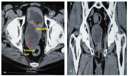
of the kidney ureter bladder region revealed a well-defined pseudo-encapsulated rounded cystic
lesion at posterior aspect of prostate in the midline measuring 6.5 cm x 5.4 cm with multiple nodules in
the wall (Figure 1).
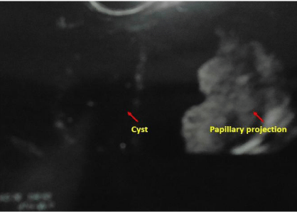
The patient underwent transrectal sonography (Figure 2) and TRUS guided biopsy.
Histopathologic examination of sections from the cyst roof showed a tumor composed of branching
papillae with central fibrovascular cores lined by pseudostratified columnar cells showing
anisonucleosis with basally located round enlarged nuclei, single prominent nucleolus and moderate
amphophilic cytoplasm. Increased mitotic activity (~8-10/10hpf) was also noted. A diagnosis of
prostatic adenocarcinoma was made. Metastatic workup (Bone scan) revealed evidence of multiple
secondary’s in the pelvic bones. In view of the stage IV disease, the patient was offered bilateral
orchidectomy plus bicalutamide 50 mg/day orally. The patient is on close follow-up.
Discussion
Most of the cysts occurring in the prostate are benign and are clinically insignificant. Benign
cysts are characterized by a smooth wall and regular outline. Prostatic abscesses are are usually
associated with clinical symptoms hence easy to diagnose. On ultrasonography prostatic abscesses
appear periurethral in location and their non-homogenous echotexture corresponds to necrotic
inflammatory debris. In contrast to this, the cysts associated with prostatic cancers have an irregular
outline with projections of solid tissue invaginations.
Malignancy associated with cystic lesions of the prostate are very
rare. Both benign and malignant prostate neoplasm’s may contain
cystic components. Multilocular prostatic cystadenoma is a rare
benign tumor that can grow to a large size causing significant lower
urinary tract symptoms. However, prostatic adenocarcinoma may
occasionally show cystic features. Other tumors of the prostate gland
that exhibit cystic components include papillary cystadenocarcinoma
and combined transitional cell/adenocarcinoma. Rarely, leiomyoma
or liposarcoma in the prostate may have cystic elements. On the MRI,
the heterogeneity of signal intensity of the cystic components and the
presence of soft-tissue elements in the lesion indicate a neoplastic cause
[4]. Aspirates from these cysts are usually haemorrhagic and contain
malignant cells. The aspirate also shows a very high concentration of
prostate specific antigen (PSA) and γ-seminoprotein [5].
The natural history of such cystic cancer of the prostate is not
well studied/known. Therefore the management of these cases do not differ from their solid counterparts as of now. The long term outcome
and biologic course of these cystic carcinomas needs further study.
Figure 1
Figure 1
a) CT scan showing a cystic mass within the prostate of the size
6.5X5.4 cm. b) The content is not clear and the cyst wall shows areas of
solid mass.
Figure 2
Figure 2
Trans-rectal ultrasonography reveals a cystic lesion in the prostate
with left lateral wall of the cyst showing solid papillary projections.
References
- Ng KL, Sathiyananthan JR, Dublin N, Razack AH and Lee G. Cystic adenocarcinoma of prostate: a case report. JUMMEC 2011; 14: 21-22.
- Walter JB, Israel MS. General pathology. 5thedn. New York. Churchill Livingstone. 1987; 386.
- Christine H. Llewellyn, Lowrey H. Holthaus. Cystic carcinoma of the prostate: Findings on Transrectal Ultrasonography. AJR. 1991; 157: 785- 786.
- Curran S, Akin O, Agildere AM, Zhang J, Hricak H, Rademaker J. Endorectal MRI of Prostatic and Periprostatic Cystic Lesions and Their Mimics. AJR 2007; 188: 1373-1379.
- Tsuji H, Hashimoto K, Katoh Y, Iguchi M. Prostatic cancer with huge cystic degeneration. Urologia Int. 1999; 62: 48-50.


