Case Report
Oral Candidiasis Turns to Oral Cancer - A Rare Clinical Presentation
Usama Kharadi*, Usama A. Kharadi, Pratik Parkarwar, Sagar Khairnar, Swati Reddy, Pradnya Arur and Tejas Kulkarni
Department of Dentistry, Oral Medicine and Radiology, P.D.U Dental College and Hospital, India
*Corresponding author: Usama Kharadi, Department of Dentistry, Oral Medicine and Radiology, P.D.U Dental College and Hospital, Solapur, India
Published: 21 Oct, 2016
Cite this article as: Kharadi U, Kharadi UA, Parkarwar P,
Khairnar S, Reddy S, Arur P, et al. Oral
Candidiasis Turns to Oral Cancer - A
Rare Clinical Presentation. Clin Oncol.
2016; 1: 1126.
Abstract
Oral carcinogens like tobacco and alcohol are well recognized risk factors associated with development oral precancerous lesions and subsequent cancer of the upper aerodigestive tract. The role of infection and chronic inflammation is also a recognized as being significant in cancer development. It has a well accepted fact that white patches of oral mucosa, which are super infected with Candida, have a greater probability of undergoing malignant transformation than those that are not infected. There are less case reports with association between chronic fungal infection and development of cancer. Here with, we report a case of chronic Candidial infection which was diagnosed and initially treated as chronic oral Candidiasis which on follow up turned into oral sqamous cell carcinoma.
Introduction
Indian subcontinent shows the highest prevalence of oral cancer among all cancers in men,
while it is the sixth most common cancer worldwide [1]. On an average possibly about 8-8.5% men
and 4-8.1% women will develop oral cancer in their lifetime in developing countries [2]. Oral cancer
is a multifactorial disease though most of the times it is preceded by some premalignant lesions
or conditions for varying length of time. Interestingly they share some etiological factor with oral
cancer, particularly the use of tobacco or other carcinogens addiction.
Infection has a considerable role in cancer development, causing approximately one in five
malignancies worldwide [3,4]. Bacteria like Helicobacter pylori, viruses like Human Papilloma
Virus (HPV), chronic hepatitis B and C infections, and herpesvirus, Epstein-Barr virus (EBV),
causes cancer due to chronic infections [5]. The literature on etiological relationship between fungal
infection and cancer are less, although for many years Candida species have been implicated in
various epithelial cancers. Candidal infection does not appear to be a risk factor for dysplastic
cervical lesions or cervical carcinoma [6] and most interest in Candida and carcinogenesis is related
to oral and esophageal carcinoma [5]. The possible association between Candida species and oral
neoplasia was first reported in the 1960s with later reports suggesting a link between the presence
of Candida albicans in the oral cavity and the development of oral squamous cell carcinoma [7]. In
actuality epithelial dysplasia improves after elimination of Candida species from infected tissue also
supports this contributory association [8].
The aim of this case report is to add in the literature of a rare case presentation of chronic oral
Candidiasis which converts into oral cancer on follow up.
Case Presentation
A 60 years female reported to department of oral medicine with a chief complaint of burning
sensation in mouth since 4-5 years. On taking detailed case history she revealed she was alright
4-5 years back when she started experiencing burning sensation in mouth. Burning sensation was
present when she uses to take spicy food which decreases on taking sweet food or sweets. Patient
was advised to take capsule Becosule for burning sensation by their relatives, but burning sensation
still persisted after taking medications once daily for few days. Now in last 15 days she visited to a
dentist for extraction of her mobile teeth in upper front region of jaw. After extraction of her teeth,
Dentist referred her to our institute for her complains regarding burning sensation in mouth and for
a white lesion on palate and tongue. No history of trauma, weakness, stress or weight loss. Patient
was asthmatic and taking inhaler for the same since last 7 to 8 years (beclomethasone dipropionate).
Patient was also hypertensive since last 3 years and on medication for the same (Tablet Amlodipine
5 mg). In general physical examination she was normal with all vital
parameters within normal limits.
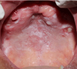
On examination of her intraoral lesions, white slightly scrapable
lesion noted involving complete palate which was also extending on
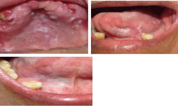
maxillary alveolar ridge in some sites (Figure 1). White non scrapable
lesion was also noted on right and left ventral surfaces of tongue
(Figure 2). There was coating on dorsal surface of tongue. Based
on her history and clinical presentation provisional diagnosis of
Pseudomembranous Candidiasis on palate and chronic hyperplastic
Candidiasis on right and left ventral surface on tongue was made.
Patient was subsequently started with topical antifungal, symptomatic
treatment and kept on follow up. After 1 month follow up lesions was
resolving on palate and ventral surfaces of tongue (Figure 3). Patient
was again called after 15 days. Lesion was still better in follow up
visits while non healing lesion was seen on maxillary anterior region
of jaw (Figure 4). Patient was given symptomatic treatment for that
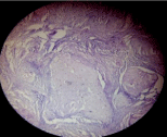
but lesion was not healing so we had opted for incisional biopsy
which was turned as well differentiated squamous cell carcinoma on
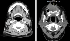
histopathologic examination (Figure 5). Patient was advised CT scan
(Figure 6) and surgical treatment was done. Patient is still on follow
up and no recurrence noted till date.
Figure 1
Figure 2
Figure 3
Figure 4
Figure 5
Figure 6
Discussion
Cancer is Latinized from Greek word ‘Karkinos’, meaning a crab,
representing how carcinoma extends its claws like a crab into the
adjacent tissues. Cancer is the second most leading cause of mortality
in economically developed countries and the third most leading
cause of death in developing countries [2]. It is estimated that more
than one million new cases are being detected annually in the Indian
subcontinent. 92-95% of all oral malignancies are oral squamous cell
carcinomas. Anyone can develop cancer; however the risk of being
diagnosed with cancer increases with age & exposure to risk factors
[9]. Longer people live the more possible it is for a sporadic mutation
to occur in their genome, leading to genetic alterations that may lead
to a malignant phenotype [9].
Oral cancer is a multifactorial disease and along with many
potentially malignant disorders, chronic oral Candidiasis is rarely
but can transfer into oral cancer. An imbalance between Candida
albicans, virulence factors and host defenses often due to specific
defects in the immune system causes Candida albicans to colonize,
penetrate, and damage host tissues [10]. There are two proposed
mechanisms by which C. albicans can invade keratinocytes. In
the first mechanism, the secretion of degradative enzymes by the
fungus, chiefly aspartic proteases that digest epithelial cell surface
components and thus allow the physical movement of hyphae into,
or between, host cells. The second proposed mechanism involve the
E-cadherin pathway in which induction of epithelial cell endocytosis,
Candida albicans stimulates keratinocytes to produce pseudopod-like
structures that encircles the fungus and depicts it into the cell in a
process [5]. Candida has capacity to induce oral cancer by directly
producing carcinogenic compounds, like nitrosamines [11]. Such
a carcinogen will attach with DNA to form adducts with bases,
phosphate residues, and/or hydrogen bonding sites which leads to
miscoding or irregularities with DNA replication [12]. The point
mutations thus induced may triggers the specific oncogenes and
instigate the development of oral cancer [5]. According to a study by
Krogh et al. [13] they proposed that some Candida species isolated
from leukoplakia lesions are capable of producing potent carcinogen
N-nitrosobenzylmethylamine (NBMA). The tubular hyphal of
Candida albicans might be important as its structure allows entrance
of precursors from saliva and a discharge of the nitrosamine product
to keratinocytes which is a potentially initiator for oral cancer [13].
Although, in a mouse model of oral carcinogenesis Dwivedi et al. [14]
found that infection with Candida albicans in itself is not capable of
inducing dysplasia or oral cancer. In another study on rat and mouse
oral squamous cell carcinoma models, Candida albicans had been
shown to act as a promoter of oral carcinogenesis where carcinogenesis
was initiated by repeated association of 4 nitroquinoline 1-oxide
(4NQO) [15]. There is a constant debate on association between
cancer and chronic inflammation since many years. There are multiple
interactions between tumor cells and stromal cells for a tumor growth
and invasion. There are many stromal events that may contribute to
carcinogenesis by initiation of specific proteolytic enzymes, which
are capable to degrade components of the basement membrane and/
or fibrous stroma [16]. According to literature Candida albicans has
ability to secrete specific proteinases, which are capable of degrading
basement membrane and extracellular matrix [17]. Candida
albicans has also been established to degrade E-cadherin, which is
a transmembrane glycoprotein, important in bonding of adjacent
keratinocytes [18]. From the above discussion we can correlate that
these findings have implication not only on the potential for Candida
albicans on tissue invasion, but also on the potential to enhance the
invasion of genetically altered epithelial cells, firstly by minimizing
the keratinocyte cohesion and then by supplementing their passage
through the basement membrane.
References
- Brad W, Neville. Oral cancer and precancerous lesions. CA Cancer J Clin. 2002; 52: 195-215.
- Garcia M, Jemal A, Ward EM, Center MM, Hao Y, Siegel RL, et al. Global Cancer Facts & Figures 2007. Atlanta, GA: American Cancer Society. 2007.
- Parkin DM. The global health burden infection-associated cancers in year. Int J Cancer. 2006; 118: 3030-3044.
- de Martel C, Franceschi S. Infections and cancer: established associations and new hypotheses. Crit Rev Oncol Hematol. 2009; 70: 183-194.
- Mohd Bakri M, Mohd Hussaini H, Rachel Holmes A, David Cannon R, Mary Rich A. Revisiting the association between candidal infection and carcinoma, particularly oral squamous cell carcinoma. J Oral Microbiol. 2010; 2: 5780.
- Engberts MK, Vermeulen CF, Verbruggen BS, van Haaften M, Boon ME, Heintz AP. Candida and squamous (pre) neoplasia of immigrants and Dutch women as established in population based cervical screening. Int J Gynecol Cancer. 2006; 16: 1596-1600.
- Gall F, Colella G, Di Onofrio V, Rossiello R, Angelillo IF, Liguori G. Candida spp. in oral cancer and oral precancerous lesions. New Microbiol. 2013; 36: 283-288.
- Williams DW, Bartie KL, Potts AJ, Wilson MJ, Fardym MJ, Lewism MA. Strain persistence of invasive Candida albicans in chronic hyperplastic candidosis that underwent malignant change. Gerodontology. 2001; 18: 73-78.
- Fedele S. Diagnostic aids in the screening of oral cancer. Head & Neck Oncology. 2009; 1: 5.
- Fidel PL Jr. Immunity to Candida. Oral Dis. 2008; 2: 69-75.
- Hooper SJ, Wilson MJ, Crean SJ. Exploring the link between microorganisms and oral cancer: a systemic review of the literature. Head Neck. 2009; 31: 1228-1239.
- Archer MC. Chemical carcinogenesis. In: Tannock IF, Hill RP, Eds. The basic science of oncology. New York: Pergamon Press. 1987; 89-105.
- Krogh P, Hald B, Holmstrup P. Possible mycological etiology of oral mucosal cancer: catalytic potential of infecting Candida albicans and other yeasts in production of N-nitrosobenzylmethylamine. Carcinogenesis. 1987; 8: 1543-1548.
- Dwivedi PP, Mallya S, Dongari-Bagtzoglou A. A novel immunocompetent murine model for Candida albicans-promoted oral epithelial dysplasia. Med Mycol. 2009; 47: 157-167.
- O’Grady JF, Reade PC. Candida albicans as a promoter of oral mucosal neoplasia. Carcinogenesis. 1992; 13: 783-786.
- Natarajan E, Saeb M, Crum CP, Woo SB, McKee PH, Rheinwald JG. Co-expression of p16 (INK4A) and laminin 5 gamma2 by microinvasive and superficial squamous cell carcinomas in vivo and by migrating wound and senescent keratinocytes in culture. Am J Pathol. 2003; 163: 477-491.
- Morschhäuser J, Virkola R, Korhonen TK, Hacker J. Degradation of human subendothelial extracellular matrix by proteinase secreting Candida albicans. FEMS Microbiol Lett. 1997; 153: 349-355.
- Pa¨rna¨nen P, Meurman JH, Samaranayake L, Virtanen I. Human oral keratinocyte E-cadherin degradation by Candida albicans and Candida glabrata. J Oral Pathol Med. 2010; 39: 275-278.


