Research Article
Video-Assisted Thoracic Surgery Efficacy in Systemic Nodal Dissection: A Single Institution Experience
Alessandro Baisi1*, Federico Raveglia2, Matilde De Simone2 and Ugo Cioffi2
1Department of Thoracic Surgery, Azienda Ospedaliera San Paolo, University of Milan, Italy
2Department of Surgery, University of Milan, Italy
*Corresponding author: Alessandro Baisi, Department of Thoracic Surgery, Azienda Ospedaliera San Paolo, University of Milan, Ospedale San Paolo Via Di Rudinì 8, 20142 Milano, Italy
Published: 12 Sep, 2016
Cite this article as: Baisi A, Raveglia F, De Simone M, Cioffi
U. Video-Assisted Thoracic Surgery
Efficacy in Systemic Nodal Dissection:
A Single Institution Experience. Clin
Oncol. 2016; 1: 1087.
Abstract
Background: According to the European Society of Thoracic Surgeons (ESTS) guidelines, systemic
nodal dissection (SND) is mandatory in pulmonary resection for non-small cell lung cancer
(NSCLC). Since VATS SND efficacy is still an uncertain issue, this is a key topic to definitively state
the oncologic effectiveness of thoracoscopy. Our study compared the number of nodes and stations
dissected and cN0-pN2 cases in VATS versus open thoracotomy.
Material and Methods: From June 2013 to December 2014, 30 patients with clinical stage I (T1-
2aN0M0) lung cancer underwent lobectomy at our thoracic surgery department. Clinical staging
was always obtained by positron emission tomography (PET). All mediastinal nodes suspected were
studied by ultrasound-guided bronchoscopy with fine needle aspiration (EBUS-FNA). Patients
were referred to VATS or open thoracotomy based on clinical general conditions and tumor
characteristics. SND was performed in both the groups.
Results: Among 30 patients who underwent lobectomy, 21 underwent open thoracotomy and 9
VATS. Four (13.3%) showed to have pN2 at definitive pathologic examination: 2 in VATS and 2
in open thoracotomy group. Mean operation time was longer in VATS than in open thoracotomy
(p=0.03).There was not significant difference between the two groups in terms of total nodes
dissected (p >0.05), mediastinal nodes dissected (p >0.05) and stations removed (p >0.05).
Conclusion: VATS SND is theoretically successful as open thoracotomy but it is technically more
demanding and more time-consuming.
Keywords: Vats; Lymphnode dissection; Non-small-cell lung cancer; Thoracotomy; Lymphnode staging
Introduction
Is widely accepted that VATS lobectomy is associated with decreased post operative pain,
shorten hospital stay, fewer post operative complications and therefore better compliance with
adjuvant chemotherapy than open lobectomy [1]. These data have prompt some thoracic surgeons
to use it routinely in early stage NSCLC.
On the other hand, many Authors still argue that VATS efficacy is uncertain in terms of
lymph node dissection and should be avoided particularly in N1 or N2 disease [2]. According to
the ESTS guidelines, it is common opinion that systemic nodal dissection (SND) is mandatory in
every pulmonary resection for NSCLC [3]. SND is recommended also in clinical N0 patients since
about 10% of them have been shown to have pathologic N2 at definitive postoperative diagnosis [4].
Therefore, the possibility to obtain SND by VATS is the key topic to definitively state the oncologic
effectiveness of thoracoscopy versus open thoracotomy.
The aim of our study was to compare the number of nodes and stations dissected in patients who
underwent lobectomy by VATS or open thoracotomy at our thoracic surgery department and to
determine if there was a significant difference in the two groups as concerns the cN0-pN2 patient’s
percentage.
Patients and Methods
From June 2013 to December 2014, 30 patients with clinical stage I (T1-2aN0M0) lung cancer
submitted to lobectomy and SND at our thoracic surgery department were retrospectively reviewed.
All data concerning surgical approach, operative time, number of stations and nodes removed and
number of cN0-N2 cases was considered. The preoperative evaluation
included cardiac assessment and pulmonary function according to
ESTS algorithm. Clinical staging was always obtained by positron
emission tomography (PET) images.
In particular, pre-operative criteria to assess the N0 stage were
the followings: 1) nodal diameter at CT< 1 cm and negative PET scan
2) nodal diameter >1 cm and negative PET scan 3) positive PET scan
without an evidence of nodal metastasis at histological examination.
All mediastinal nodes suspected were studied by ultrasound-guided
bronchoscopy with fine needle aspiration (EBUS-FNA).
Patients were classified in two groups based on the surgical
approach. Group 1 consisting of 9 patients that underwent VATS and
group 2 consisting of 21 patients that underwent open thoracotomy.
Patients were referred to VATS or open thoracotomy by the same
thoracic surgeon team based on clinical general conditions and tumor
characteristics (size, position, etc.). However, surgical approach was
never determined based on nodal status since all patients have already
been classified N0.
All procedures were performed by the same Team of two
surgeons. We always used the Copenhagen technique [1]. Patents
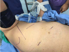
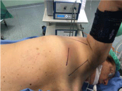
were placed in posterolateral decubitus. VATS procedures were done
by two centimeter incisions and a minithoracotomy with the surgeon
positioned anteriorly: 1) the first incision, for the camera port, was
performed on the anterior axillary line in the 7th intercostal space 2)
the second incision in the 5th intercostal space on the posterior axillary
line 3) lastly a 4-5 cm minithoracotomy in the anterior 5th intercostal
space. The ribs spreader was never used. Open thoracotomy was
always a posterolateral incision in the 5th intercostal space with the
surgeons positioned at the patients’ back. We always performed
systemic nodal dissection in both the groups at the end of lobectomy.
On the right side the stations dissected were 2,3,4,7,8,9 and 11, on
the left side 4,5,6,7,8,9,10 and 11. Subcarinal, pulmonary vein,
paraesophageal and hilar nodes were resected singularly. Paratracheal
dissection was performed removing the fatty soft tissue including 4L,
2R and 4R nodes. Nodal dissection was initially performed using
a bipolar forceps in the two groups then we have introduced the
harmonic scalpel.
Data were expressed as mean +/- standard deviation (SD).
Difference between groups with continuous variables were assessed
by the X2 test or Fischer’s exact test.
Statistical significance was accepted as p <0.05.
Figure 1
Figure 2
Results
30 patients underwent lobectomy for stage I NSCLC. Twenty-one
underwent open thoracotomy and 9 VATS. Four (13.3%) showed to
have pN2 at definitive pathologic examination (cN0-pN2). Two were
in the VATS group and 2 in the open thoracotomy group. Among
these patients, 1 presented mediastinal nodal positivity at PET and
was determined to be free of metastasis by EBUS-FNA. Both the
groups underwent SND.
The two groups were similar as concerning age, sex, histology,
pulmonary function (p >0.05). Comparing the operative features, the
mean operation time in VATS was longer than in open thoracotomy
with a statistical significance (p=0.03), while intraoperative blood loss,
chest tube duration and hospital staying were equivalent (p>0.05).
There was no significant difference between the two groups in
terms of total nodes dissected (16.2 +/- 5.4 per patients in the VATS
group vs. 18.8 +/- 7.8 in the open thoracotomy group, p >0.05),
mediastinal nodes dissected (10.5 +/- 5 per patients in the VATS
group vs. 12.5 +/- 5.5 in the open thoracotomy group, p >0.05) and
stations removed (6.8 +/- 1.6 per patients in the VATS group vs. 8
+/- 2.5 in the open thoracotomy group, p >0.05). However, the overall
number of nodes and stations removed in open thoracotomy group
was slightly higher. Despite, our data did not allow to statistically
analyzing every single station, number 7 and 4L had less nodes
removed in the VATS group. The most of patients with cN0-pN2
nodes presented a singular station and singular node metastasis in
both the groups (7/8 and 10/11 cases respectively). The analysis of
cN0-pN2 nodes in either group revealed that none of PET positive
nodes previously studied by EBUS was metastatic.
There were 5 cases of conversion to open thoracotomy that were
enrolled in the group 2. All conversions were performed based on
technical difficulties as incomplete fissure or severe adhesions. There
were no complications or in hospital deaths in either group.
Discussion
Accurate clinical tumor staging is essential in the management
of lung cancer since the indication to surgery with curative intent is
restricted only to cases with local disease (stage I and II). The N status
assessment is probably the most challenging issue in clinical staging.
PET scan has improved the accuracy of the non invasive procedures
but is still affected by a remarkable percentage of false negative cases
as reported by Kim and Co-workers [5]. False negative nodes are
related in the most of cases to the impossibility in detecting micrometastasis.
On the contrary, mediastinal nodes enlargement at CT
scan or PET/CT positivity without a real malignant involvement, are
frequent in smokers or BPCO patients. Based on these evidences, it
is inevitable that postoperative N upstaging could happen as reported
in literature by several Authors [6,7]. Therefore preoperative nodal
biopsy is recommended in case of suspected malignant involvement.
In summary, invasive procedures can be omitted in patients
with peripheral tumors and negative mediastinal (PET) images,
whereas PET positive mediastinal findings should always be cytohistologically
confirmed. Transbronchial needle aspiration (TBNA),
ultrasound-guided bronchoscopy with fine needle aspiration (EBUSFNA)
and endoscopic esophageal ultrasound-guided fine needle
aspiration (EUS-FNA) are techniques that provide cyto-histological
diagnosis and are minimally invasive. Their specificity is high but the
negative predictive value is low. Mediastinoscopy is more invasive
but is the most accurate method. Nowadays, EBUS-FNA and the
mediastinoscopy are routinely used to preoperatively perform
mediastinal nodal staging.
Despite these non-invasive and invasive techniques, Certfolio
and Colleagues have showed that about 10% of clinical N0 become
N2 after complete nodal dissection [6].
Based on the evidence that clinical nodal staging is a challenging
issue, the ESTS guidelines [3] recommend to always perform an
intraoperative complete nodal dissection to resect the all the possible
undetected pathologic nodes also in N0 patients. The goal is to
obtain the most accurate postoperative nodal status assessment and
determine the real prognosis and the best postoperative treatment.
It is widely accepted that open thoracotomy allows an effective
complete nodal dissection. On the contrary, the remaining concern
about VATS lobectomy is the completeness of nodal dissection versus
open thoracotomy. These concerns are about the followings items:
the total number of nodes dissected, the number of stations sampled
and tissue fragmentation. In particular, some nodal stations as 7 and
4L are considered very demanding to be reached during thoracoscopy
and tissue fragmentation could be associated with seeding risks.
Few studies have faced this important issue. At the beginning
of the VATS era, it was generally accepted that nodal dissection by
thoracoscopy was not as successful as open thoracotomy. Therefore,
frozen section of mediastinal nodes was advocated in order to convert
to open thoracotomy in case of a pathologic response. Then, when
VATS technique became more familiar to surgeons, some Authors
were able to show that the number of stations and nodes dissected in
VATS versus thoracotomy were similar [8]. New surgical techniques
have also been developed to do the dissection of the most demanding
nodal station. For instance, Baste has recently described an anterior
approach to dissect nodes number 7 after an inferior left lobectomy
[9]. However, serious doubts are persistent in several Authors.
Our data concerning the number of nodes resected by open
thoracotomy and cN0-pN2 cases are similar to those reported in
literature. The comparison between VATS and open thoracotomy
showed that the two techniques were equivalent. These results,
according with the latest literature [10,11], seem to confirm the
oncologic efficacy of VATS lobectomy.
The most important bias of our paper is the small population
enrolled. Moreover, it is a retrospective study therefore patients have
been not randomized. However, we underline that surgical approach
has never been chosen based on nodes characteristics. These could
have influenced the results.
In our opinion, thoracoscopic approach to mediastinal nodes is
a controversial technique. Its most interesting advantage is the closer
view of the anatomical structures that allows a safer and comfortable
dissection. On the contrary, the most disturbing characteristic is the
discomfortable access to some deep stations as the number 7 and 4L.
Conclusion
VATS nodal dissection is theoretically equivalent to open thoracotomy but it is technically more demanding and probably more time consuming. In wider terms, VATS approach to pulmonary lobectomy and SND is one of the most specialized procedures in thoracic surgery and should be reserved to high specialized institutions.
References
- Yan TD, Cao C, D'Amico TA, Demmy TL, He J, Hansen H, et al. International VATS Lobectomy Consensus Group. Video-assisted thoracoscopic surgery lobectomy at 20 years: a consensus statement. Eur J Cardiothorac Surg. 2014; 45: 633-639.
- Merritt RE, Hoang CD, Shrager JB. Lymph node evaluation achieved by open lobectomy compared with thoracoscopic lobectomy for N0 lung cancer. Ann Thorac Surg. 2013; 96: 1171-1177.
- De Leyn P, Dooms C, Kuzdzal J, Lardinois D, Passlick B, Rami-Porta R, et al. Revised ESTS guidelines for preoperative mediastinal lymph node staging for non-small-cell lung cancer. Eur J Cardiothorac Surg. 2014; 45: 787-798.
- Wang S, Zhou W, Zhang H, Zhao M, Chen X. Feasibility and long-term efficacy of video-assisted thoracic surgery for unexpected pathologic N2 disease in non-small cell lung cancer. Ann Thorac Med. 2013; 8: 170-175.
- Pak K, Park S, Cheon GJ, Kang KW, Kim IJ, Lee DS, et al. Update on nodal staging in non-small cell lung cancer with integrated positron emission tomography/computed tomography: a meta-analysis. Ann Nucl Med. 2015; 2: 6.
- Cerfolio RJ, Bryant AS, Minnich DJ. Complete thoracic mediastinal lymphadenectomy leads to a higher rate of pathologically proven N2 disease in patients with non-small cell lung cancer. Ann Thorac Surg. 2012; 94: 902-906.
- Baisi A, Raveglia F, De Simone M, Cioffi U. The role of video-assisted thoracic surgery lobectomy in unexpected N2 cases. Ann Thorac Surg. 2014; 97: 1125.
- Boffa DJ, Kosinski AS, Paul S, Mitchell JD, Onaitis M. Lymph node evaluation by open or video-assisted approaches in 11,500 anatomic lung cancer resections. Ann Thorac Surg. 2012; 94: 347-353.
- Baste JM, Haddad L, Melki J, Peillon C. Anterior subcarinal node dissection on the left side using video thoracoscopy: an easier technique. Ann Thorac Surg. 2015; 99: e99-e101.
- Wang W, Yin W, Shao W, Jiang G, Wang Q, Liu L, et al. Comparative study of systematic thoracoscopic lymphadenectomy and conventional thoracotomy in resectable non-small cell lung cancer. J Thorac Dis 2014; 6: 45-51.
- Baisi A, Rizzi A, Raveglia F, Cioffi U. Video-assisted thoracic surgery is effective in systemic lymph node dissection. Eur J Cardiothorac Surg 2013; 44: 966.


