Clinical Image
Renal Cell Carcinoma with Parieto-Occipital Bone Metastasis
Mittal V*, Rupala KK, Ahmad J and Suryavanshi M
Department of Urology, Kidney Transplant and Robotics, India
*Corresponding author: Varun Mittal, Department of Urology, Kidney Transplant and Robotics, Medanta, Medicity Gurgaon, Haryana, India
Published: 07 Sep, 2016
Cite this article as: Mittal V, Rupala KK, Ahmad J,
Suryavanshi M. Renal Cell Carcinoma
with Parieto-Occipital Bone Metastasis.
Clin Oncol. 2016; 1: 1075.
Abstract
One third patients with renal cell carcinoma (RCC) have metastatic disease at presentation. Eight to 15% patients present with metastases in head and neck region. Skull bone metastasis is a very rare presentation. Herein we report a case of 56 year male RCC with parieto-occipital bone metastasis. He underwent radiotherapy for skull lesion and cytoreductive nephrectomy for right renal mass. Metastatic RCC is a diagnostic dilemma especially when there is no clue in clinical presentation. An unusual vascular osteolytic lesion in head and neck in middle-aged person should be dealt with a high index of suspicion for RCC.
Clinical Image
Renal cell carcinoma (RCC) constitutes about 3% of adult malignancies and 90-95 % of neoplasm
arising from the kidney [1]. Renal cell carcinoma patients are mostly asymptomatic at time of
diagnosis and are detected incidentally. Approximately one third of patients present with distant
metastasis. Frequently metastasis is seen in lung, bone, adrenal, liver, and contralateral kidney [2].
Head and neck is involved in around 8-15% of metastatic renal tumor and sites include paranasal
sinuses, larynx, mandible, temporal bones and thyroid gland [2,3]. There are very few reported cases
of renal cell carcinoma presenting as parieto-occipiatal bone metastasis.
Herein we present a case of a 56 years old gentleman who presented with painless, progressively
increasing right sided scalp mass for 6 months. On examination it was large around 10 X 10cm, non
tender parieto-occipiatal swelling leading to right ear deformity, (Figure 1). Fine needle aspiration
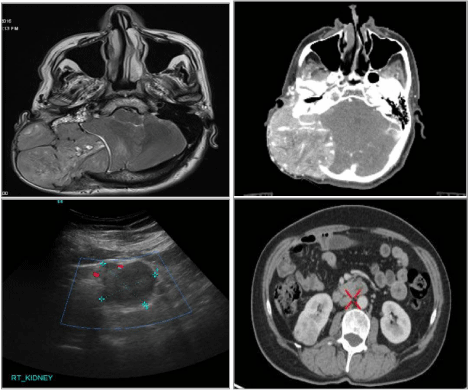
cytology revealed metastatic epithelioid malignancy. Magnetic resonance and computerized
tomography imaging of head revealed a large lobulated expansile intensely enhancing mass lesion
centered in scalp over right occipital and retromastoid region causing destruction of underlying
occipital bone with intracranial and extradural extension. During evaluation for the primary lesion
ultrasound abdomen revealed approximately 4.8 X 4.2cm exophytic mass lesion arising from lower
pole of right kidney, (Figure 2). Patient was referred to urology from neurosurgery. Whole body
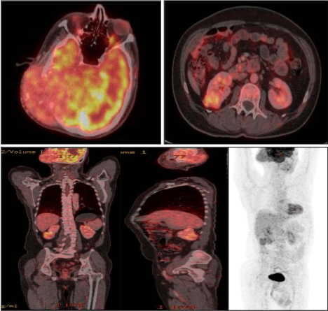
positron emission with contrast enhanced computerized tomography scan (PET-CECT) done for
staging of primary disease revealed a FDG avid large mass (9.1 X 6.6 X 7.7cm) with intracranial and
extracranial extension with involvement of right parietal and occipital bones with midline shift,
FDG avid subcarinal lymph node (1.9 X 3.6 cm) and FDG avid 3.5 X 3.7 X 4.2cm mass arising
from lower pole of right kidney, (Figure 2 and 3). Findings were consistent with metastatic renal
cell carcinoma. Scalp lesion was subjected to Intensity Modulated Radiotherapy in a dose of 40
Gray in 10 Fractions. He underwent right laparoscopic cytoreductive
nephrectomy. His post operative period was uneventful discharged
on post operative day 2. Histopathology report revealed pT1b,
Fuhrman's nuclear grade 3, clear cell carcinoma, with free margins.
On immunohistochemistry it was CK 7: negative. Patient is doing
well at 6 months follow up. Scalp swelling has become somewhat soft
with overlying alopecia of scalp.
Renal cell carcinoma rarely metastasize to head and neck bones
but in any patient with skull bone metastasis and unknown primary,
high index of suspicion of RCCs should be kept. The prognosis of
such patient is extremely poor with 1 year survival rate of 20% [3].
Treatment intent is usually cytoreductive, immunomodulation and
palliative.
Figure 1
Figure 1
Large, 10 x 10 cm, non tender parieto-occipiatal swelling over right side of scalp displacing right ear.
Figure 2
Figure 2
Magnetic resonance and computerized tomography imaging of head depicting a large lobulated expansile intensely enhancing mass lesion centered in
scalp over right occipital and retromastoid region causing destruction of underlying occipital bone with intracranial and extradural extension. Ultrasound abdomen
revealed approximately 4.8 X 4.2 cm exophytic mass lesion arising from lower pole of right kidney which was confirmed on PET- CECT abdomen. (PET-CECT,
positron emission with contrast enhanced computerized tomography scan).
Figure 3
Figure 3
Whole body PET-CECT showing a FDG avid large mass (9.1 X 6.6 X 7.7 cm) with intracranial and extracranial extension with involvement of right parietal
and occipital bones with midline shift, FDG avid subcarinal lymph node (1.9 X 3.6 cm) and FDG avid 3.5 X 3.7 X 4.2 cm mass arising from lower pole of right kidney.
(PET-CECT, positron emission with contrast enhanced.
References
- Sepúlveda I, Platin E, Klaassen R, Spencer ML, García C, Alarcón R, et al. Skull base clear cell carcinoma, metastasis of renal primary tumor: a case report and literature review. Case Rep Oncol. 2013; 6: 416-423.
- Jallu A, Latoo M, Pampori R. Rare case of renal cell carcinoma with mandibular swelling as primary presentation. Case Rep Urol. 2013; 806192.
- De Vos C, Gerard JM, Decat M, Gersdorff M. Metastatic renal cell carcinoma to the temporal bone: case report. B-ENT. 2005; 1: 43-46.


