Case Report
Misdiagnosing Hepatic Inflammatory Pseudotumor as Hepatocellular Carcinoma: A Case Report
Lei Y1, Bin Z1, Ying-he Q1, Ning-jia S1, Xin-yuan L2, Wan Yee L1,3, LAI, Eric CH1,3, Yong-jie Z1*
1Second Department of Biliary Surgery, Eastern Hepatobiliary Surgery Hospital, China
2Department of Pathology, Eastern Hepatobiliary Surgery Hospital, China
3The Chinese University of Hong Kong, Prince of Wales Hospital, Hong Kong
*Corresponding author: Zhang, Yong-jie, Second Department of Biliary Surgery, Eastern Hepatobiliary Surgery Hospital, Shanghai, #225 Changhai Road, Shanghai, People's Republic of China
Published: 06 Sep, 2016
Cite this article as: Lei Y, Bin Z, Ying-he Q, Ning-jia S, Xinyuan
L, Wan Yee L, et al. Misdiagnosing
Hepatic Inflammatory Pseudotumor
as Hepatocellular Carcinoma: A Case
Report. Clin Oncol. 2016; 1: 1072.
Abstract
A 61-year-old Chinese male was found to have a lesion in the left liver on routine body check-up.
Laboratory tests revealed no abnormalities except for a rise in C-reactive protein (CRP). Computed
tomography (CT) showed features suggestive of hepatocellular carcinoma (HCC). The patient
underwent liver IVb segmentectomy and cholecystectomy. Histopathology showed features of
hepatic inflammatory pseudotumor. The CRP decreased to close to normal on postoperative day 9.
The patient was discharged home well on postoperative day 11.
Keywords: Hepatic inflammatory pseudotumor; Hepatocellular carcinoma; Hepatectomy; Cholecystectomy
Introduction
Inflammatory pseudotumor (IPT) is rare and it is characterized by chronic infiltration of
inflammatory cells with areas of fibrosis [1]. Brunn first described pulmonary IPT in 1939 [2]. IPT
occurs usually in the lung and rarely in the parotid gland, pleura, stomach, ovary and liver. Hepatic
inflammatory pseudotumor (HIPT) was first described by Pack and Baker in 1953 [3]. So far, more
than 200 cases of HIPT have been reported in the medical literature [4]. It is a challenge to diagnose
HIPT because of its rarity, indistinctive clinical manifestations and non-typical radiological features.
It is easy to misdiagnose HIPT as other benign or malignant hepatic tumors.
We herein report one patient with HIPT which mimicked as hepatocellular carcinoma (HCC).
Case Presentation
A 61-year-old Chinese male was found to have a lesion in the left liver on routine medical checkup.
There was no complaints and physical examination showed no abnormalities.
Laboratory tests showed normal liver function, hemoglobin, white cells and platelet counts.
However, the C-reaction protein (CRP) was raised to 60.4 mg/L. The hepatitis B e antibody and
hepatitis B core antibody were positive while the hepatitis B surface antigen and anti-hepatitis C
virus were both negative. The α-fetoprotein, carcinoembryonic antigen, carbohydrate antigen19-9,
carbohydrate antigen125, carbohydrate antigen153 and des-γ-carboxy prothrombin were all
normal.
Chest radiography revealed no abnormalities. An ultrasound revealed a hypoechoic mass, 68 x
50 mm, in segment IV of liver. The lesion had irregular borders and heterogeneous internal echo.
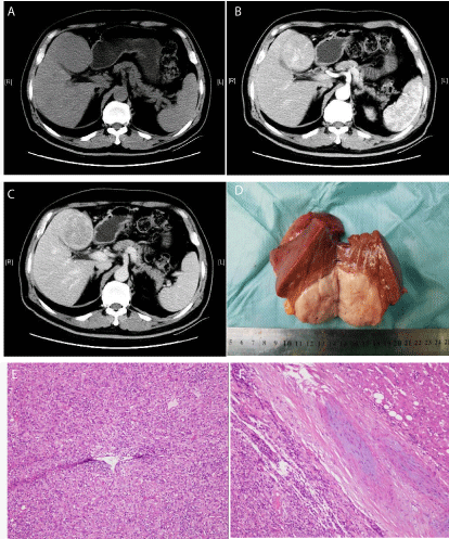
Contrast-enhanced computed tomography (CT) revealed a quasi-circular, low density mass of 7 x
5 x 5 cm, with indistinct borders and heterogeneous density (Figure A). The lesion was enhanced
unevenly in the arterial phase (Figure B) with contrast washout in the venous phase (Figure C).
The patient underwent IVb segmentectomy and cholecystectomy. Grossly the mass was quasicircular,
solid, 7 x 5 x 5 cm in size, and was located in segment IVb of the liver. The lesion was close
to the gallbladder. The liver showed no evidence of cirrhosis. There was no enlarged lymph node.
There were no invasion into adjacent organs, extrahepatic metastases and peritoneal seedlings.
The operation took 145 minutes. The time of the Pringle’s maneuver was 12 minutes and the
blood loss was 100 ml.
The surgical specimen showed a fishlike and uniform mass (Figure D) and histopathology
revealed a hepatic inflammatory pseudotumor. The lesion contained dense collagen fiber bundles
which were disorderly arranged, and dispersed with fusiform
myofibroblasts / fibroblasts without any atypia. It contained diffuse
infiltration of chronic inflammatory cells, mainly plasma cells and
lymphocytes (Figure E). A thickened wall and luminal stenosis in
the hepatic vein led to the formation of non-inflammatory venous
occlusion (Figure F).
The CRP decreased to 11.2 mg/L on postoperative day 9. The
patient was discharged home well on postoperative day 11.
Figure
Figure
Abdominal computed tomography revealed a large, solid mass (7
cm in size) in segment IV of liver. Contrast-enhanced computed tomography
(CT) imaging revealed a quasi-circular, low density mass of 7 x 5 x 5 cm
with unclear borders and heterogeneous density (Figures A). The lesion
was enhanced unevenly in the arterial phase (Figures B) and washout in
the venous phase (Figures C). The surgical specimen showed a fishlike
and uniform mass (Figures D) and histopathological examination revealed
a hepatic inflammatory pseudotumor. The lesion contained dense collagen
fiber bundles which were disorderly arranged, dispersed with fusiform
myofibroblasts / fibroblasts without any atypia. The lesion contained
diffuse infiltration of chronic inflammatory cells, mainly plasma cells and
lymphocytes (E). The thickened wall and luminal stenosis in hepatic vein led
to the formation of non-inflammatory venous occlusion (F).
Discussion
HIPT is rare. The etiology and pathogenesis of this disease is
not clear. Some researchers have demonstrated that the etiology and
pathogenesis of HIPT may be related to factors such as infection,
immune response, radiation, and chemotherapy [5].
HIPT can occur at any age. The ratio of male/female in adult
HIPT ranged from 1:1-3.5:1 [6]. Patients with HIPT often present
with atypical clinical symptoms such as abdominal pain, fever and
weight loss, and they often have no history of hepatitis and cirrhosis.
There is no clear correlation with hepatitis B viral infection. The
liver function and tumor markers are usually normal or just slightly
elevated. Laboratory tests may reveal an inflammatory process, with
leukocytosis or increased CRP. Radiological features are atypical.
HIPT is often shown to have hypo- or isodensity on CT, hypo- or
isointense on T1WI and hyper- or isointense on T2WI on magnetic
resonance imaging. Without histopathological studies, it is difficult
to arrive at a correct diagnosis and HIPT is easy to be misdiagnosed
as other liver tumors.
Our patient was completely asymptomatic. The HBeAb(+) and
HBcAb(+) revealed a past history of HBV infection. CT showed a
liver mass with low density which enhanced in the arterial phase. We
clinically misdiagnosed this lesion as hepatocellular carcinoma before
surgery because of the laboratory and medical imaging findings.
Percutaneous needle biopsy was not done on this patient because
of the potential risk of tumor seeding along the needle tract. There
have been reports on malignant conversion of HIPT [7].
This patient had elevated CRP before surgery. CRP is an acutephase
protein created by the liver. There have been reports which
showed that an increase in CRP was associated with severity of
inflammation in several liver diseases, such as chronic hepatitis B&C
and HCC [8-11]. The average level of CRP in HCC has been reported
to be 30.78±15.17 mg/l (normal 0.78±1.07 mg/l) [12]. Several case
reports have also revealed that the CRP in HIPT was raised [13, 14].
In our patient, the preoperative CRP of 60.4 mg/L and a postoperative
CRP level of 11.2 mg/L demonstrated that the HIPT was associated
with the raised CRP.
In summary, a patient with HIPT who presented with features
mimicking HCC was reported. A preoperatively raised CRP was the
only hint which suggested that our patient might had HIPT instead
of HCC.
References
- Ahn KS, Kang KJ, Kim YH, Lim TJ, Jung HR, Kang YN, et al. Inflammatory pseudotumors mimicking intrahepatic cholangiocarcinoma of the liver; IgG4-positivity and its clinical significance. J Hepatobiliary Pancreat Sci. 2012: 19; 405-412.
- Umiker WO, L Iverson. Postinflammatory tumors of the lung; report of four cases simulating xanthoma, fibroma, or plasma cell tumor. J Thorac Surg. 1954: 28; 55-63.
- Pack GT, HW Baker. Total right hepatic lobectomy; report of a case. Ann Surg. 1953. 138: 253-258.
- Akatsu T, Wakabayashi G, Tanimoto A, Kameyama K, Kitajima M. Inflammatory pseudotumor of the liver masquerading as hepatocellular carcinoma after a hepatitis B virus infection: Report of a case. Surg Today. 2006: 36; 1028-1031.
- Coffin CM1, Watterson J, Priest JR, Dehner LP. Extrapulmonary inflammatory myofibroblastic tumor (inflammatory pseudotumor). A clinicopathologic and immunohistochemical study of 84 cases. Am J Surg Pathol. 1995: 19; 859-872.
- Shek TW, Ng IO, Chan KW. Inflammatory pseudotumor of the liver. Report of four cases and review of the literature. Am J Surg Pathol. 1993: 17; 231-238.
- Pecorella I, Memeo L, Trombetta G, de Quarto A, de Simone P, et al. Inflammatory pseudotumour of the liver--evidence for malignant transformation. Pathol Res Pract. 1999. 195: 115-120.
- Andreozzi P, Viscogliosi G, Colella F, Subic M, Cipriani E, Marigliano B, et al. [Predictors of liver fibrosis in patients with non-alcoholic fatty liver disease. The role of metabolic syndrome, insulin-resistance and inflammation]. Recenti Prog Med. 2012: 103; 570-574.
- Ma LN, Liu XY, Luo X, Hu YC, Liu SW, Tang YY, et al. Serum high-sensitivity C-reactive protein are associated with HBV replication, liver damage and fibrosis in patients with chronic hepatitis B. Hepatogastroenterology. 2015: 62; 368-372.
- Sjowall C, Cardell K, Boström EA, Bokarewa MI, Enocsson H, Ekstedt M, et al. High prevalence of autoantibodies to C-reactive protein in patients with chronic hepatitis C infection: association with liver fibrosis and portal inflammation. Hum Immunol. 2012: 73: 382-388.
- Nishikawa H, Arimoto A, Wakasa T, Kita R, Kimura T, Osaki Y. Pre-treatment C-reactive protein as a prognostic factor for recurrence after surgical resection of hepatocellular carcinoma. Anticancer Res. 2013: 33; 1181-1188.
- Liu XY, Ma LN, Yan TT, Lu ZH, Tang YY, Luo X, et al. Combined detection of liver stiffness and C-reactive protein in patients with hepatitis B virus-related liver cirrhosis, with and without hepatocellular carcinoma. Mol Clin Oncol. 2016: 4; 587-590.
- Kruth J, Michaely H, Trunk M, Niedergethmann M, Rupf AK, Krämer BK, et al. A rare case of fever of unknown origin: inflammatory myofibroblastic tumor of the liver. Case report and review of the literature. Acta Gastroenterol Belg. 2012: 75; 448-453.
- Al-Hussaini H, Azouz H, Abu-Zaid A. Hepatic inflammatory pseudotumor presenting in an 8-year-old boy: A case report and review of literature. World J Gastroenterol. 2015: 21; 8730-8.