Case Report
Gallbladder Melanoma Presenting with Acute RUQ Pain
Frederico F*, Weatherly B, Weiss E, Carroll B and Earl T
Department of Radiology, The University of Mississippi Medical Center, USA
*Corresponding author: Frederico F. Souza, Department of Radiology, The University of Mississippi Medical Center, USA
Published: 29 Jul, 2016
Cite this article as: Frederico F, Weatherly B, Weiss E,
Carroll B, Earl T. Gallbladder Melanoma
Presenting with Acute RUQ Pain. Clin
Oncol. 2016; 1: 1039.
Abstract
We present a case of a 37 year old male with multiple co-morbidities including hemophilia A, HIV, and chronic hepatitis C who presented with progressively worsening right upper quadrant pain and intermittent nausea. Ultrasound demonstrated polypoid echogenic lesions along the gallbladder wall with flow on power Doppler. CT scan of the abdomen was subsequently performed and demonstrated enhancement of the gallbladder lesions. An MRI of the abdomen was also obtained and showed a dominant T1 hyper intense enhancing mass involving the gallbladder lumen, multiple enlarged lymph nodes and enhancing hepatic lesions promoting peripheral capsular retraction. The patient subsequently underwent diagnostic cholecystectomy and liver biopsy. Surgical pathology of the gallbladder was diagnostic for malignant melanoma, with liver biopsies also diagnostic for metastatic malignant melanoma.
Introduction
Melanoma is an aggressive neoplasm which, while only accounting for approximately 1% of reported cancers in the United States, is increasing in prevalence [1]. Melanoma typically arises from preexisting benign nevi with metastases potentially involving any organ, though the most common sites of metastases are the lungs, liver, and central nervous system. Melanoma spreads by direct extension as well as lymphatic and hematogenous dissemination. While low grade melanoma can have a 100% cure rate with wide excision, the majority of lesions are detected late with an overall mortality rate of 30-40% [2]. A well-known potential site of melanoma metastasis is the gallbladder, with melanoma accounting for more than 50% of gallbladder metastases. Gallbladder metastases are also discovered at autopsy in 15% of patients who die of disseminated melanoma [1,2]. We present a case of a 37-year-old with gallbladder melanoma who underwent extensive imaging workup.
Case Report
The patient is a 37-year-old male who presented with progressively worsening rightupper
quadrant pain and intermittent nausea. The patient was afebrile with a blood pressure of 138/88
mmHg. On physical exam, he had right upper quadrant and right lateral abdominal tenderness to
palpation, with an absent Murphy’s sign. His past medical history was significant for hemophilia A,
HIV, and chronic hepatitis C.
A routine chest radiograph was normal. Subsequently, a dedicated gallbladder ultrasound
was recommended for further evaluation and demonstrated polypoid echogenic lesions along the
gallbladder wall (Figure 1A) with flow on power Doppler, reflecting internal vascularity (Figure
1B). Given the concern for possible gallbladder neoplasm, a subsequent CT of the abdomen
and pelviswith and without intravenous contrast (Triple phase scan - Liver mass protocol) was
performed (Figure 2A and B) which showed enhancement of the gallbladder lesions. Of note, no
liver lesions were identified on CT imaging. An MRI of the abdomen was obtained two days later
to assess potential involvement of the adjacent liver parenchyma. The MRI showeda dominant
T1hyperintense enhancing mass involving the gallbladder lumen (Figure 3A and B) and multiple
enlarged lymph nodes in the upper abdomen which also demonstratedT1 hyper intensity.
Additionally, there were numerous ill-defined T1 hyperintenseenhancing lesions throughout the
liver promoting peripheral capsular retraction (Figure 3A). The patient subsequently underwent IR
percutaneous gallbladder biopsy. The pathology results were inconclusive and the patient eventually
underwent diagnostic cholecystectomy and liver biopsy. Surgical pathology of the gallbladder was
diagnostic for malignant melanoma, with liver biopsies also diagnostic for metastatic malignant
melanoma. As no other focus was identified as a result of extensive imaging and physical exam
evaluation, the patient was diagnosed with primary malignant melanoma of the gallbladder. During
admission, the patient subsequently died of cardio respiratory complications.
Figure 1A
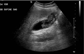
Figure 1A
Ultrasound B mode in the right upper quadrant demonstrates a
echogenic mass in the body and fundus of the gallbladder.
Figure 1B
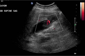
Figure 1B
Power Doppler demonstrating flow/vascularity in the central
portion of the mass.
Figure 2A
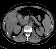
Figure 2A
CT scan of the abdomen obtained before the administration of
IV contrast demonstrates a hiperdense area in the fundus of the gallbladder.
Discussion
Melanoma of the GI tract is relatively uncommon; antemortem diagnosis is made in only 1.5–4.4% of all patients with melanoma, however melanoma represents about one third of all metastatic disease of the GI tract and is found in 58% of postmortem specimens from patients with melanoma [3]. While melanoma has a clinical course that can involve diffuse metastases to many sites, [1,4] both primary and metastatic melanoma to the gallbladder are extremely rare, with only 20 cases of primary melanoma and 60 cases of metastatic melanoma to the gallbladder reported as of 2008. However, melanoma is the most common tumor to metastasize to the gallbladder, accounting for 30-60% of gallbladder metastases [1]. There have been several case reports of primary melanoma in the gallbladder [5-8] as well as metastatic melanoma in the gallbladder [1,2,9]. Gallbladder melanoma is most often found in patients with disseminated disease. It has been reported that 15% of patients who died of malignant melanoma had gallbladder metastasis at autopsy [1,2]. Clinically, however, gallbladder involvement by melanoma has been relatively occult, with symptoms seldom presenting [1], making our case of a patient who presented with symptoms suggestive of cholecystitis rare. Two case reports in the surgical and pathology literature have described gallbladder melanoma presenting with cholecystitis-like symptoms [5,8,9]. Our isolated presentation of gallbladder melanoma is, therefore, seldom seen among cases of this rare entity. During our patient’s diagnostic course, imaging of this rare case of gallbladder melanoma was obtained utilizing ultrasound, CT, and MRI. In particular, following concern raised for a neoplastic etiology due to the polypoid gallbladder lesions demonstrating both internal vascularity on US and enhancement on CT, the presence of T1 hyperintensity in both the gallbladder and metastatic liver lesions on MRI raised concern for melanoma as the likely etiology. This paramagnetic characteristic of melanin has been thought to be due to the presence of free radicals known to occur in melanin [10,11]. Others have theorized, however, that scavenging of paramagnetic metals by melanin accounts for their short T1 times [12]. The pathologic distinction between primary and secondary melanoma of the gallbladder is challenging. The primary site of disease may be elusive, as cutaneous melanomas may be small and regress over time, and thus may be difficult to detect. Due to the migration of melaninproducing cells from the neural crest to endodermal derivatives during embryogenesis, there are some melanocytes in gallbladder mucosa, making development of primary gallbladder melanoma a possibility [1]. In addition to a solitary nature, pathologic characteristics considered requisite to make a diagnosis of primary gallbladder melanoma include a papillary or polypoid configuration arising from the gallbladder mucosa and the presence of junctional activity [5]. The true existence of primary gallbladder melanoma, however, has been questioned [6]. A final consideration in our presented case is the development of melanoma in an HIV patient. A study in Lancet in 2007 demonstrated a similarincidence of melanoma in patients with HIV and in immunosupreessed transplant recipients [13], suggesting that immunosuppression, rather than HIV infection itself ,is associated with incidence of melanoma.
Figure 2B
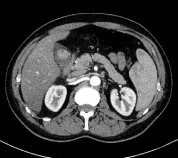
Figure 2B
Axial CT scan of the abdomen after administration of intravenous
contrast demonstrates that the mass has avid enhancement.
Figure 3
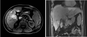
Figure 3
Axial and coronal T1 Fat Sat pre contrast imaging of the upper
abdomen demonstrates multiple hyperintense T1 lesions in the liver,
hyperintense T1 adenopathy in the porta-caval region and hyperintese
gallbladder mass (better seen on the coronal plane).
Conclusion
We present a case of primary or secondary melanoma of the gallbladder. This case is unique in the literature given not only its isolated presentation, but also its manifestation presenting with cholecystitis-like symptoms in a patient with HIV.
References
- Hamodat M, Alhumidi A. Malignant melanoma of the gall bladder, a case report and literature review. Internet J Path. 2008; 9.
- Guida M, Cramarossa A, Gentile A. Metastatic malignant melanoma of the gallbladder: a case report and review of the literature. Melanoma Res. 2002; 12: 619-625.
- Reinhardt MJ, Joe AY, Jaeger U. Diagnostic performance of whole body dual modality 18F-FDG PET/CT imaging for N- and M-staging of malignant melanoma: experience with 250 consecutive patients. J Clin Oncol. 2006; 24: 1178-1187.
- Dasgupta T, Brasfield R. Metastatic melanoma. A clinicopathological study. Cancer. 1964; 17: 1323-1339.
- Heath DI, Womack C. Primary malignant melanoma of the gall bladder. J ClinPathol. 1988; 41: 1073-1077.
- Safioleas M, Agapitos E, Kontzoglou, K. Primary melanoma of the gallbladder. Does it exist? Report of a case and review of the literature. World J Gastroenterol. 2006; 12: 4259-4261.
- Velez AF, Penetrante RB, Spellman JE Jr. Malignant melanoma of the gallbladder: report of a case and review of the literature. Am Surg, 1995; 61: 1095-1098.
- HaskaracaMF, Ozsoy M, Özsan I. Primary malignant melanoma of the gallbladder: a case report and review of the literature. Case Reports in Surg. 2012; Art ID 693547.
- Henriques CQ. A case of secondary melanoma of the gall-bladder presenting as acute cholecystitis. Brit J Surg. 1955; 42: 663-665.
- Aime S, Botta M, Ermondi G. Paramagnetic water proton relaxation enhancement : from contrast agents in MRI to reagents for quantitative « in vitro » assays. Magn Reson Imaging. 1992; 10: 849-854.
- Atlas S, Braffman B, LoBrutto R. Human malignant melanomas with varying degrees of melanin content in nude mice : MR imaging, histopathology, and electron paramagnetic resonance. J Compute Assist Tomogr. 1990;14 : 547-554.
- Enochs WS, Petherick P, Bogdanova A. Paramagnetic metal scavenging by melanin: MR imaging. Radiology. 1997; 204: 417-423.
- Grulich AE, van Leeuwen MT, Falster MO. Incidence of cancers in people with HIV/AIDS compared with immunosuppressed transplant recipients: a meta-analysis. Lancet. 2007; 370: 59-67.