Case Report
A 25 Year-Old Male with Cerebellar Mass: Lhermitte-Duclos Disease
Ulger S1*, Demirci H2, Çeltikçi E2, Kurt G2, Kilic D1, Karakus E3, Helvaci A3, Ucar M4 and Uluoglu O3
1Department of Radiation Oncology, Gazi University, Turkey
2Department of Neurosurgery, Gazi University, Turkey
3Department of Pathology, Gazi University, Turkey
4Department of Radiology, Gazi University, Turkey
*Corresponding author: Sukran Ulger, Department of Radiation Oncology, Gazi University Hospital, 06500 Besevler- Ankara, Turkey
Published: 18 Jul 2016
Cite this article as: Ulger S, Demirci H, Çeltikçi E, Kurt G,
Kilic D, Karakus E, et al. A 25 Year-Old
Male with Cerebellar Mass: Lhermitte-
Duclos Disease. Clin Oncol. 2016; 1:
1023.
Abstract
We report a rare case of dysplastic cerebellar gangliocytoma also called as Lhermitte-Duclos Disease
located in the cerebellum in a young male without any neurological signs. Morphologically dysplastic
neurons and immunohistochemical staining with neuronal and glial markers was all diagnostic on
pathology. Also a non-enhancing cerebellar hemispheric mass with a striated pattern of hyper-, and
isointensity on T2-weighted images is classical findings on brain MRI.
MRI findings of the lesion is
Highlights
1. Dysplastic cerebellar gangliocytoma is an uncommon hamartomatous disorder.
2. The classical MRI findings of the lesion is a non-enhancing cerebellar hemispheric mass
with a striated pattern of hyper-, and isointensity on T2-weighted images.
3. Keep in mind in younger patients without any neurological signs and hamartomatous
lesions.
Keywords: Lhermitte duclos disease; Cerebellar mass; Gangliocytoma
Introduction
Dysplastic cerebellar gangliocytoma is an uncommon hamartomatous disorder which was first described by Lhermitte and Duclos in 1920 as a cerebellar ganglion cell tumor. Since that, although more than ninety years passed, approximately a hundred new cases have been reported in the literature [1]. So we decided to report our patient to contribute to the literature and for the brain pathologist to not to forget the diagnosis as a differential diagnosis on brain tumor specimens and keep in mind in younger patients without any neurological signs which is more rare.
Case Presentation
A 25-year-old man referred to our neurosurgery department with pain on his neck and back.
The neurological examination and physical examination were completely normal. Magnetic
resonance (MR) imaging of the brain was performed. A mass lesion of abnormal signal intensity
was detected. Axial T2 weighted MR image (Figure 1A) showed heterogeneous hyperintensity
with striated appearance of the left cerebellum with mass effect in the fourth ventricle and regions
with centrally cystic appearance. Contrast-enhanced axial T1-weighted image (Figure 1B) showed
very subtle enhancement within the lesion. Edema and mass effect was not detected. The patient
underwent a gross total resection of tumor.
No neurological disorders were detected postoperatively and have been followed without any
recurrence since the last 6 months. The genetic evaluation showed constitutional karyotype (46,XY).
Brain sectioning from cerebellum revealed, dispersed glial and neuronal elements. The neurons
had dysplastic morphologic changes such as cytomegaly, multinucleation and Nissl bodies
located perimembranous (Figure 2A). There was classification and ectatic vascular structures on
histopathological evaluation. There was no necrosis and mitosis was not detected microscopically.
The proliferation index of the tumor was less than 1% after examination with ki-67 staining.
The conventional and immunohistochemical staining results of
synaptophysin (Figure 2B), CD34 and neurofilaments dye was
positive on dysplastic neurons which have bi-nuclear pleomorphic
morphology and also immunohistochemical staining was positive on
the extensions of dysplastic neurons.
The immunohistochemical staining with glial fibrillary acidic
protein (GFAP) was strongly positive on cellular intermediate
filaments in neoplastic glial component and slightly positive as a
perinuclear rim on glial cells.
Histochemical examination with reticuline dye was also positive
on vascular bed and intermediate filaments.
The histopathological diagnosis was Dysplastic Cerebellar
Gangliocytoma: Lhermitte-Duclos Disease.
Figure 1
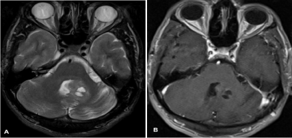
Figure 1
Axial T2 weighted MR image (A) shows heterogeneous hyperintensity with striated appearance of the left cerebellum with mass effect in the fourth
ventricle and regions with centrally cystic appearance. Contrast-enhanced axial T1-weighted image (B) shows very subtle enhancement within the lesion.
Figure 2
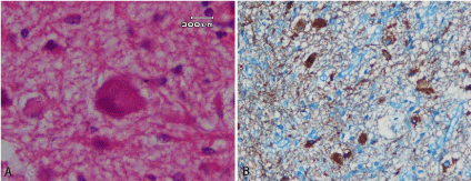
Figure 2
Nissl bodies located perimembranous on dysplastic neurons (haematoxylin and eosin staining, original magnification X1000) (A), Displastic neurons
show consistent cytoplasmic staining for synaptophysin (immunhistochemistry, original magnification X400) (B).
Discussion
Lhermitte-Duclos Disease is classified as a WHO grade 1
tumor. The pathogenesis of LDD is not clearly defined. It can
occur sporadically or in familial form. Cowden syndrome which is
known as an autosomal dominant hereditary malignancy syndrome
characterized by multiple hamartomas, mucocutaneous lesions
(trichilemmomas, facial papules, mucosal papillomas, and acral
keratosis), and a high incidence of other neoplasms such as breast,
thyroid, genitourinary, and endometrial cancers, is closely associated
with LDD which is seen in approximately 40% of LDD patients [2].
Cowden syndrome and LDD are considered as a single
phakomatosis with autosomal dominant inheritance, which is caused
by a mutation in the PTEN suppressor oncogene [3]. There was no
genetic abnormality and no mutation in our patients’ gene analysis.
The lesions frequently cause progressive mass effects in the
posterior fossa and are commonly associated with symptoms of
intracranial hypertension and cerebellar dysfunction. The most
common presentation is in the third and fourth decades of life.
Dysplastic gangliocytoma presents rarer in younger patients [3]. Our
patient differs from the other cases with both no neurological signs
and symptoms at presentation and younger age.
In recent years, MR imaging achieved major importance
as a diagnostic tool in the preoperative evaluation of dysplastic
gangliocytoma. The classical MRI findings of the lesion is described
as non-enhancing cerebellar hemispheric mass with a striated
pattern of hyper-, and isointensity on T2-weighted images. Contrast
enhancements have been reported in LDD lesions rarely. Also the
post-contrast T2 images showed slight contrast enhancing in our
patient. For long-term follow-up MRI is recommended [4].
Tumor resection by a suboccipital approach is the surgical
procedure generally performed. The patients are usually reported
as successfully relieved of symptoms by surgical excision. The longterm
survival improves from 2.5 to 11 years with surgical treatment.
Although total resection is not possible generally due to the invasion
of the brainstem. Nonetheless ‘watch and wait’ is a reasonable
approach for the patients without neurological symptoms due to the
benign nature of the tumor [4]. Our patient had severe neck pain and
our surgeons were doubtful about the relationship between the pain
and the mass. So they decided to operate the patient despite he had no
neurological signs. Also the neck pain resolved after surgery.
Radiation therapy has also been reported in some patients with
Lhermitte-Duclos disease, but unfortunately the results seem not to
be satisfactory [5]. The outcome in unoperated patients is usually
poor due to the mass effect of the tumor.
Conclusion
Lhermitte-Duclos disease can be easily diagnosed with neuroimaging modalities and neuro-pathologic evaluation with a care-full work-up, can be treated by surgery with an improved survival nowadays and must be kept in mind in asymptomatic and neurologically normal and younger patients. It is important to report all the patients diagnosed LDD to clarify the clinical outcome of the disease.
References
- Shinagare AB. Patil NK, Sorte SZ. Case 144: Dysplastic Cerebellar Gangliocytoma (Lhermitte-Duclos Disease). Radiology. 2009; 251: 298-303.
- Wells GB, Lasner TM, Yousem DM, Zager EL. Lhermitte-Duclos disease and Cowden's syndrome in an adolescent patient. Case report. J Neurosurg. 1994; 81: 133-136.
- Capone Mori A, Hoeltzenbein M, Poetsch M, Schneider JF, Brandner S, Boltshauser E. Lhermitte-Duclos disease in 3 children: a clinical long-term observation. Neuropediatrics. 2003; 34: 30-35.
- Yağci-Küpeli B, Oguz KK, Bilen MA, Yalçin B, Akalan N, Büyükpamukçu M. An unusual cause of posterior fossa mass: Lhermitte-Duclos disease. J Neurol Sci. 2010; 290: 138-141.
- Prestor B. Dysplastic gangliocytoma of the cerebellum (Lhermitte-Duclos disease). J Clini Neurosci. 2006; 13: 877–881.